3NR6 | pdb_00003nr6

Crystal structure of xenotropic murine leukemia virus-related virus (XMRV) protease


Experimental Data Snapshot

  • Method: X-RAY DIFFRACTION
  • Resolution: 1.97 Å
  • R-Value Free: 
    0.233 (Depositor), 0.248 (DCC) 
  • R-Value Work: 
    0.196 (Depositor), 0.207 (DCC) 
  • R-Value Observed: 
    0.197 (Depositor) 

Starting Model: in silico
View more details

wwPDB Validation 3D Report Full Report

Validation slider image for 3NR6

This is version 1.4 of the entry. See complete history

Literature

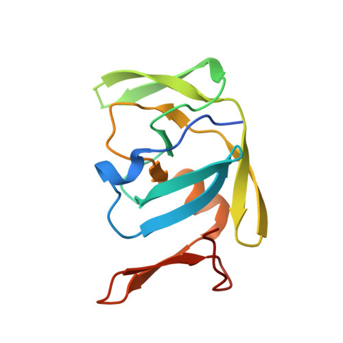
Crystal structure of XMRV protease differs from the structures of other retropepsins.

Li, M.Dimaio, F.Zhou, D.Gustchina, A.Lubkowski, J.Dauter, Z.Baker, D.Wlodawer, A.

(2011) Nat Struct Mol Biol 18: 227-229

  • DOI: https://doi.org/10.1038/nsmb.1964
  • Primary Citation Related Structures: 
    3NR6

  • PubMed Abstract: 

    Using energy and density guided Rosetta refinement to improve molecular replacement, we determined the crystal structure of the protease encoded by xenotropic murine leukemia virus-related virus (XMRV). Despite overall similarity of XMRV protease to other retropepsins, the topology of its dimer interface more closely resembles those of the monomeric, pepsin-like enzymes. Thus, XMRV protease may represent a distinct branch of the aspartic protease family.


  • Organizational Affiliation
    • Protein Structure Section, Macromolecular Crystallography Laboratory, National Cancer Institute at Frederick, Frederick, Maryland, USA.

Macromolecule Content 

  • Total Structure Weight: 28.82 kDa 
  • Atom Count: 1,883 
  • Modeled Residue Count: 234 
  • Deposited Residue Count: 264 
  • Unique protein chains: 1

Macromolecules

Find similar proteins by:|  3D Structure
Entity ID: 1
MoleculeChains  Sequence LengthOrganismDetailsImage
Protease p14
A, B
132Xenotropic MuLV-related virus VP62Mutation(s): 0 
Gene Names: gag-pol
EC: 3.4.23
UniProt
Find proteins for A1Z651 (Xenotropic MuLV-related virus (isolate VP62))
Explore A1Z651 
Go to UniProtKB:  A1Z651
Entity Groups
Sequence Clusters30% Identity50% Identity70% Identity90% Identity95% Identity100% Identity
UniProt GroupA1Z651
Sequence Annotations
Expand
Reference Sequence

Experimental Data & Validation

Experimental Data

  • Method: X-RAY DIFFRACTION
  • Resolution: 1.97 Å
  • R-Value Free:  0.233 (Depositor), 0.248 (DCC) 
  • R-Value Work:  0.196 (Depositor), 0.207 (DCC) 
  • R-Value Observed: 0.197 (Depositor) 
Space Group: P 4 2 2
Unit Cell:
Length ( Å )Angle ( ˚ )
a = 63.915α = 90
b = 63.915β = 90
c = 106.509γ = 90
Software Package:
Software NamePurpose
MAR345data collection
PHASERphasing
Rosettamodel building
REFMACrefinement
HKL-3000data reduction
HKL-3000data scaling
Rosettaphasing

Structure Validation

View Full Validation Report


Entry History 

Deposition Data

Revision History  (Full details and data files)

  • Version 1.0: 2011-02-02
    Type: Initial release
  • Version 1.1: 2011-07-13
    Changes: Version format compliance
  • Version 1.2: 2017-11-08
    Changes: Refinement description
  • Version 1.3: 2023-12-27
    Changes: Data collection, Database references, Derived calculations
  • Version 1.4: 2024-04-03
    Changes: Refinement description