3NL6

The Crystal Structure of Candida glabrata THI6, a Bifunctional Enzyme involved in Thiamin Biosyhthesis of Eukaryotes


Experimental Data Snapshot

  • Method: X-RAY DIFFRACTION
  • Resolution: 2.61 Å
  • R-Value Free: 0.271 
  • R-Value Work: 0.221 
  • R-Value Observed: 0.223 

wwPDB Validation   3D Report Full Report


Ligand Structure Quality Assessment 


This is version 1.3 of the entry. See complete history


Literature

Domain Organization in Candida glabrata THI6, a Bifunctional Enzyme Required for Thiamin Biosynthesis in Eukaryotes .

Paul, D.Chatterjee, A.Begley, T.P.Ealick, S.E.

(2010) Biochemistry 49: 9922-9934

  • DOI: https://doi.org/10.1021/bi101008u
  • Primary Citation of Related Structures:  
    3NL2, 3NL3, 3NL5, 3NL6, 3NM1, 3NM3

  • PubMed Abstract: 

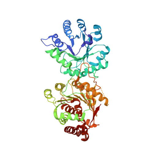
    THI6 is a bifunctional enzyme found in the thiamin biosynthetic pathway in eukaryotes. The N-terminal domain of THI6 catalyzes the ligation of the thiamin thiazole and pyrimidine moieties to form thiamin phosphate, and the C-terminal domain catalyzes the phosphorylation of 4-methyl-5-hydroxyethylthiazole in a salvage pathway. In prokaryotes, thiamin phosphate synthase and 4-methyl-5-hydroxyethylthiazole kinase are separate gene products. Here we report the first crystal structure of a eukaryotic THI6 along with several complexes that characterize the active sites responsible for the two chemical reactions. THI6 from Candida glabrata is a homohexamer in which the six protomers form a cage-like structure. Each protomer is composed of two domains, which are structurally homologous to their monofunctional bacterial counterparts. Two loop regions not found in the bacterial enzymes provide interactions between the two domains. The structures of different protein-ligand complexes define the thiazole and ATP binding sites of the 4-methyl-5-hydroxyethylthiazole kinase domain and the thiazole phosphate and 4-amino-5-hydroxymethyl-2-methylpyrimidine pyrophosphate binding sites of the thiamin phosphate synthase domain. Our structural studies reveal that the active sites of the two domains are 40 Å apart and are not connected by an obvious channel. Biochemical studies show 4-methyl-5-hydroxyethylthiazole phosphate is a substrate for THI6; however, adenosine diphospho-5β-ethyl-4-methylthiazole-2-carboxylic acid, the product of THI4, is not a substrate for THI6. This suggests that an unidentified enzyme is necessary to produce the substrate for THI6 from the THI4 product.


  • Organizational Affiliation

    Department of Chemistry and Chemical Biology, Cornell University, Ithaca, New York 14853, United States.


Macromolecules
Find similar proteins by:  (by identity cutoff)  |  3D Structure
Entity ID: 1
MoleculeChains Sequence LengthOrganismDetailsImage
Thiamine biosynthetic bifunctional enzyme
A, B, C
540Nakaseomyces glabratusMutation(s): 0 
Gene Names: CAGL0E05808gTHI6
EC: 2.5.1.3 (PDB Primary Data), 2.7.1.50 (PDB Primary Data)
UniProt
Find proteins for Q6FV03 (Candida glabrata (strain ATCC 2001 / BCRC 20586 / JCM 3761 / NBRC 0622 / NRRL Y-65 / CBS 138))
Explore Q6FV03 
Go to UniProtKB:  Q6FV03
Entity Groups  
Sequence Clusters30% Identity50% Identity70% Identity90% Identity95% Identity100% Identity
UniProt GroupQ6FV03
Sequence Annotations
Expand
  • Reference Sequence
Small Molecules
Ligands 3 Unique
IDChains Name / Formula / InChI Key2D Diagram3D Interactions
ACP
Query on ACP

Download Ideal Coordinates CCD File 
F [auth A],
I [auth B],
L [auth C]
PHOSPHOMETHYLPHOSPHONIC ACID ADENYLATE ESTER
C11 H18 N5 O12 P3
UFZTZBNSLXELAL-IOSLPCCCSA-N
TPS
Query on TPS

Download Ideal Coordinates CCD File 
E [auth A],
H [auth B],
K [auth C]
THIAMIN PHOSPHATE
C12 H18 N4 O4 P S
HZSAJDVWZRBGIF-UHFFFAOYSA-O
MG
Query on MG

Download Ideal Coordinates CCD File 
D [auth A],
G [auth B],
J [auth C]
MAGNESIUM ION
Mg
JLVVSXFLKOJNIY-UHFFFAOYSA-N
Experimental Data & Validation

Experimental Data

  • Method: X-RAY DIFFRACTION
  • Resolution: 2.61 Å
  • R-Value Free: 0.271 
  • R-Value Work: 0.221 
  • R-Value Observed: 0.223 
  • Space Group: C 1 2 1
Unit Cell:
Length ( Å )Angle ( ˚ )
a = 163.173α = 90
b = 153.528β = 117.99
c = 109.618γ = 90
Software Package:
Software NamePurpose
PHENIXrefinement
PDB_EXTRACTdata extraction

Structure Validation

View Full Validation Report


Ligand Structure Quality Assessment 


Entry History 

Deposition Data

Revision History  (Full details and data files)

  • Version 1.0: 2010-11-10
    Type: Initial release
  • Version 1.1: 2011-07-13
    Changes: Version format compliance
  • Version 1.2: 2013-05-29
    Changes: Other
  • Version 1.3: 2024-02-21
    Changes: Data collection, Database references, Derived calculations