3NKN | pdb_00003nkn

Crystal structure of mouse autotaxin in complex with 14:0-LPA


Experimental Data Snapshot

  • Method: X-RAY DIFFRACTION
  • Resolution: 1.80 Å
  • R-Value Free: 
    0.234 (Depositor), 0.228 (DCC) 
  • R-Value Work: 
    0.184 (Depositor), 0.179 (DCC) 
  • R-Value Observed: 
    0.187 (Depositor) 

Starting Model: experimental
View more details

wwPDB Validation 3D Report Full Report

Validation slider image for 3NKN

Ligand Structure Quality Assessment 


This is version 2.2 of the entry. See complete history

Literature

Crystal structure of autotaxin and insight into GPCR activation by lipid mediators

Nishimasu, H.Okudaira, S.Hama, K.Mihara, E.Dohmae, N.Inoue, A.Ishitani, R.Takagi, J.Aoki, J.Nureki, O.

(2011) Nat Struct Mol Biol 18: 205-212

  • DOI: https://doi.org/10.1038/nsmb.1998
  • Primary Citation Related Structures: 
    3NKM, 3NKN, 3NKO, 3NKP, 3NKQ, 3NKR

  • PubMed Abstract: 

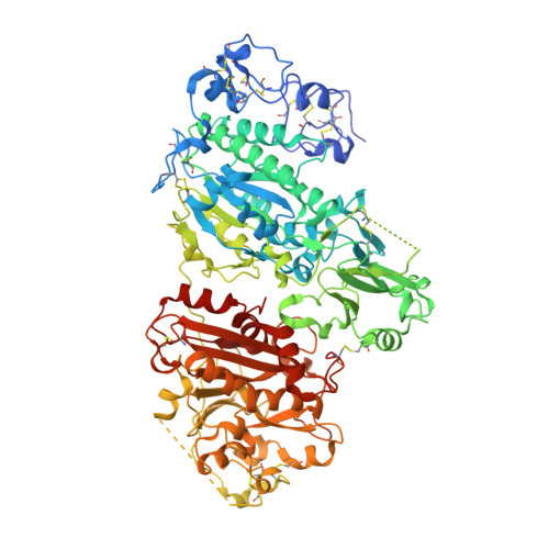
    Autotaxin (ATX, also known as Enpp2) is a secreted lysophospholipase D that hydrolyzes lysophosphatidylcholine to generate lysophosphatidic acid (LPA), a lipid mediator that activates G protein-coupled receptors to evoke various cellular responses. Here, we report the crystal structures of mouse ATX alone and in complex with LPAs with different acyl-chain lengths and saturations. These structures reveal that the multidomain architecture helps to maintain the structural rigidity of the lipid-binding pocket, which accommodates the respective LPA molecules in distinct conformations. They indicate that a loop region in the catalytic domain is a major determinant for the substrate specificity of the Enpp family enzymes. Furthermore, along with biochemical and biological data, these structures suggest that the produced LPAs are delivered from the active site to cognate G protein-coupled receptors through a hydrophobic channel.


  • Organizational Affiliation
    • Department of Biophysics and Biochemistry, Graduate School of Science, The University of Tokyo, Tokyo, Japan.

Macromolecule Content 

  • Total Structure Weight: 100.1 kDa 
  • Atom Count: 7,189 
  • Modeled Residue Count: 782 
  • Deposited Residue Count: 831 
  • Unique protein chains: 1

Macromolecules

Find similar proteins by:|  3D Structure
Entity ID: 1
MoleculeChains  Sequence LengthOrganismDetailsImage
Ectonucleotide pyrophosphatase/phosphodiesterase family member 2831Mus musculusMutation(s): 0 
Gene Names: Enpp2Npps2Pdnp2
EC: 3.1.4.39 (PDB Primary Data), 3.1.4.4 (UniProt)
UniProt
Find proteins for Q9R1E6 (Mus musculus)
Explore Q9R1E6 
Go to UniProtKB:  Q9R1E6
Entity Groups
Sequence Clusters30% Identity50% Identity70% Identity90% Identity95% Identity100% Identity
UniProt GroupQ9R1E6
Glycosylation
Glycosylation Sites: 3Go to GlyGen: Q9R1E6-1
Sequence Annotations
Expand
Reference Sequence

Oligosaccharides

Help  
Entity ID: 2
MoleculeChains Length2D Diagram GlycosylationD Interactions
2-acetamido-2-deoxy-beta-D-glucopyranose-(1-4)-2-acetamido-2-deoxy-beta-D-glucopyranose
B, D
2N-Glycosylation
Glycosylation Resources
GlyTouCan: G42666HT
GlyCosmos: G42666HT
GlyGen: G42666HT
Entity ID: 3
MoleculeChains Length2D Diagram GlycosylationD Interactions
alpha-D-mannopyranose-(1-2)-alpha-D-mannopyranose-(1-3)-alpha-D-mannopyranose-(1-6)-beta-D-mannopyranose-(1-4)-2-acetamido-2-deoxy-beta-D-glucopyranose-(1-4)-2-acetamido-2-deoxy-beta-D-glucopyranose
C
6N-Glycosylation
Glycosylation Resources
GlyTouCan: G47410OF
GlyCosmos: G47410OF
GlyGen: G47410OF

Small Molecules

Ligands 7 Unique
IDChains Name / Formula / InChI Key2D Diagram3D Interactions
NKN

Query on NKN


Download:Ideal Coordinates CCD File
J [auth A],
K [auth A]
(2R)-2-hydroxy-3-(phosphonooxy)propyl tetradecanoate
C17 H35 O7 P
FAZBDRGXCKPVJU-MRXNPFEDSA-N
ZN

Query on ZN


Download:Ideal Coordinates CCD File
E [auth A],
F [auth A]
ZINC ION
Zn
PTFCDOFLOPIGGS-UHFFFAOYSA-N
EDO

Query on EDO


Download:Ideal Coordinates CCD File
AA [auth A]
BA [auth A]
CA [auth A]
DA [auth A]
EA [auth A]
AA [auth A],
BA [auth A],
CA [auth A],
DA [auth A],
EA [auth A],
FA [auth A],
GA [auth A],
HA [auth A],
N [auth A],
O [auth A],
P [auth A],
Q [auth A],
R [auth A],
S [auth A],
T [auth A],
U [auth A],
V [auth A],
W [auth A],
X [auth A],
Y [auth A],
Z [auth A]
1,2-ETHANEDIOL
C2 H6 O2
LYCAIKOWRPUZTN-UHFFFAOYSA-N
SCN

Query on SCN


Download:Ideal Coordinates CCD File
L [auth A],
M [auth A]
THIOCYANATE ION
C N S
ZMZDMBWJUHKJPS-UHFFFAOYSA-M
CA

Query on CA


Download:Ideal Coordinates CCD File
G [auth A]CALCIUM ION
Ca
BHPQYMZQTOCNFJ-UHFFFAOYSA-N
K

Query on K


Download:Ideal Coordinates CCD File
I [auth A]POTASSIUM ION
K
NPYPAHLBTDXSSS-UHFFFAOYSA-N
NA

Query on NA


Download:Ideal Coordinates CCD File
H [auth A]SODIUM ION
Na
FKNQFGJONOIPTF-UHFFFAOYSA-N

Experimental Data & Validation

Experimental Data

  • Method: X-RAY DIFFRACTION
  • Resolution: 1.80 Å
  • R-Value Free:  0.234 (Depositor), 0.228 (DCC) 
  • R-Value Work:  0.184 (Depositor), 0.179 (DCC) 
  • R-Value Observed: 0.187 (Depositor) 
Space Group: P 1 21 1
Unit Cell:
Length ( Å )Angle ( ˚ )
a = 61.58α = 90
b = 94.617β = 94.86
c = 75.372γ = 90
Software Package:
Software NamePurpose
DENZOdata reduction
SCALEPACKdata scaling
PHENIXrefinement
PDB_EXTRACTdata extraction
ADSCdata collection
HKL-2000data reduction

Structure Validation

View Full Validation Report


Ligand Structure Quality Assessment 


Entry History 

Deposition Data

Revision History  (Full details and data files)

  • Version 1.0: 2011-01-19
    Type: Initial release
  • Version 1.1: 2011-07-13
    Changes: Version format compliance
  • Version 1.2: 2014-02-19
    Changes: Database references
  • Version 1.3: 2017-11-08
    Changes: Refinement description
  • Version 2.0: 2020-07-29
    Type: Remediation
    Reason: Carbohydrate remediation
    Changes: Advisory, Atomic model, Data collection, Database references, Derived calculations, Structure summary
  • Version 2.1: 2023-11-01
    Changes: Data collection, Database references, Refinement description, Structure summary
  • Version 2.2: 2024-11-13
    Changes: Structure summary