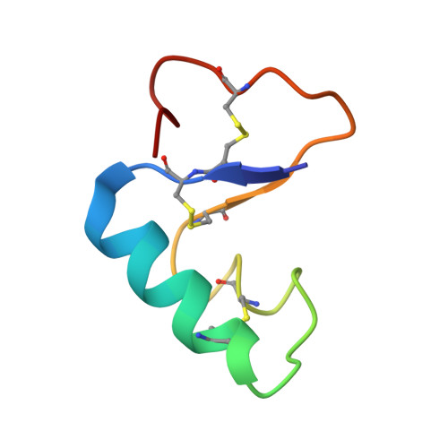
Crystal structure of small protein crambin at 0.48 A resolution
Schmidt, A., Teeter, M., Weckert, E., Lamzin, V.S.(2011) Acta Crystallogr Sect F Struct Biol Cryst Commun 67: 424-429
- PubMed: 21505232 Search on PubMedSearch on PubMed Central
- DOI: https://doi.org/10.1107/S1744309110052607
- Primary Citation Related Structures:
3NIR - PubMed Abstract:
With the development of highly brilliant and extremely intense synchrotron X-ray sources, extreme high-resolution limits for biological samples are now becoming attainable. Here, a study is presented that sets the record in crystallographic resolution for a biological macromolecule. The structure of the small protein crambin was determined to 0.48 Å resolution on the PETRA II ring before its conversion to a dedicated synchrotron-radiation source. The results reveal a wealth of details in electron density and demonstrate the possibilities that are potentially offered by a high-energy source. The question now arises as to what the true limits are in terms of what can be seen at such high resolution. From what can be extrapolated from the results using crystals of crambin, this limit would be at approximately 0.40 Å, which approaches that for smaller compounds.
- EMBL Hamburg, c/o DESY, Notkestrasse 85, D-22607 Hamburg, Germany. andrea@embl-hamburg.de
Organizational Affiliation: