


Crystal structure of the TPR domain of kinesin light chain 1
Tong, Y., Tempel, W., Shen, L., Shen, Y., Nedyalkova, L., Arrowsmith, C.H., Edwards, A.M., Bountra, C., Weigelt, J., Bochkarev, A., Park, H.To be published.
Experimental Data Snapshot
Starting Model: experimental
View more details
wwPDB Validation 3D Report Full Report

Entity ID: 1 | |||||
|---|---|---|---|---|---|
| Molecule | Chains | Sequence Length | Organism | Details | Image |
| Kinesin light chain 1 | 311 | Homo sapiens | Mutation(s): 0 Gene Names: KLC1, KLC, KNS2 | 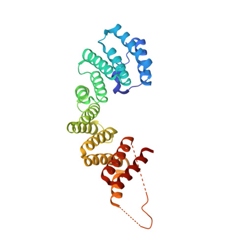 | |
UniProt & NIH Common Fund Data Resources | |||||
PHAROS: Q07866 GTEx: ENSG00000126214 | |||||
Entity Groups | |||||
| Sequence Clusters | 30% Identity50% Identity70% Identity90% Identity95% Identity100% Identity | ||||
| UniProt Group | Q07866 | ||||
Sequence AnnotationsExpand | |||||
Reference Sequence | |||||
| Length ( Å ) | Angle ( ˚ ) |
|---|---|
| a = 74.685 | α = 90 |
| b = 74.685 | β = 90 |
| c = 156.168 | γ = 120 |
| Software Name | Purpose |
|---|---|
| DENZO | data reduction |
| SCALEPACK | data scaling |
| PHASER | phasing |
| PDB_EXTRACT | data extraction |
| HKL-2000 | data reduction |
| HKL-2000 | data scaling |
| BUSTER | refinement |


