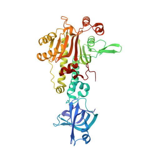
Crystal structure of aspartyl-tRNA synthetase from Pyrococcus kodakaraensis KOD: archaeon specificity and catalytic mechanism of adenylate formation
Schmitt, E., Moulinier, L., Fujiwara, S., Imanaka, T., Thierry, J.C., Moras, D.(1998) EMBO J 17: 5227-5237
- PubMed: 9724658 Search on PubMedSearch on PubMed Central
- DOI: https://doi.org/10.1093/emboj/17.17.5227
- Primary Citation Related Structures:
1B8A, 3NEL, 3NEM, 3NEN - PubMed Abstract:
The crystal structure of aspartyl-tRNA synthetase (AspRS) from Pyrococcus kodakaraensis was solved at 1.9 A resolution. The sequence and three-dimensional structure of the catalytic domain are highly homologous to those of eukaryotic AspRSs. In contrast, the N-terminal domain, whose function is to bind the tRNA anticodon, is more similar to that of eubacterial enzymes. Its structure explains the unique property of archaeal AspRSs of accommodating both tRNAAsp and tRNAAsn. Soaking the apo-enzyme crystals with ATP and aspartic acid both separately and together allows the adenylate formation to be followed. Due to the asymmetry of the dimeric enzyme in the crystalline state, different steps of the reaction could be visualized within the same crystal. Four different states of the aspartic acid activation reaction could thus be characterized, revealing the functional correlation of the observed conformational changes. The binding of the amino acid substrate induces movement of two invariant loops which secure the position of the peptidyl moiety for adenylate formation. An unambiguous spatial and functional assignment of three magnesium ion cofactors can be made. This study shows the important role of residues present in both archaeal and eukaryotic AspRSs, but absent from the eubacterial enzymes.
- Laboratoire de Biologie Structurale, Institut de Génétique et de Biologie Moléculaire et Cellulaire, CNRS/INSERM/ULP, 1 rue Laurent Fries, BP163, 67404 Illkirch Cédex, C.U. de Strasbourg, France.
Organizational Affiliation: