3NEK | pdb_00003nek

Crystal structure of a nitrogen repressor-like protein MJ0159 from Methanococcus jannaschii


Experimental Data Snapshot

  • Method: X-RAY DIFFRACTION
  • Resolution: 2.50 Å
  • R-Value Free: 
    0.277 (Depositor), 0.248 (DCC) 
  • R-Value Work: 
    0.246 (Depositor), 0.250 (DCC) 
  • R-Value Observed: 
    0.247 (Depositor) 

wwPDB Validation 3D Report Full Report

Validation slider image for 3NEK

This is version 1.5 of the entry. See complete history

Literature

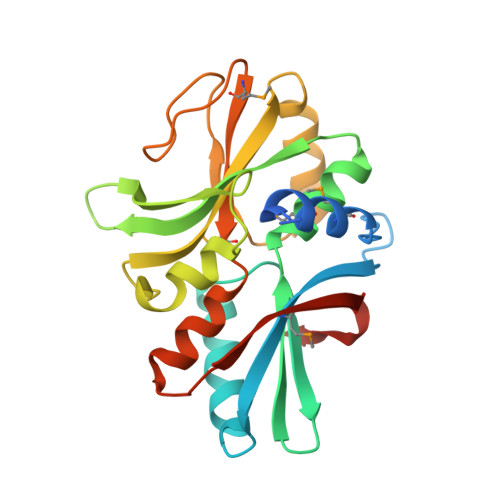
Structural underpinnings of nitrogen regulation by the prototypical nitrogen-responsive transcriptional factor NrpR.

Wisedchaisri, G.Dranow, D.M.Lie, T.J.Bonanno, J.B.Patskovsky, Y.Ozyurt, S.A.Sauder, J.M.Almo, S.C.Wasserman, S.R.Burley, S.K.Leigh, J.A.Gonen, T.

(2010) Structure 18: 1512-1521

  • DOI: https://doi.org/10.1016/j.str.2010.08.014
  • Primary Citation Related Structures: 
    3NEK

  • PubMed Abstract: 

    Plants and microorganisms reduce environmental inorganic nitrogen to ammonium, which then enters various metabolic pathways solely via conversion of 2-oxoglutarate (2OG) to glutamate and glutamine. Cellular 2OG concentrations increase during nitrogen starvation. We recently identified a family of 2OG-sensing proteins--the nitrogen regulatory protein NrpR--that bind DNA and repress transcription of nitrogen assimilation genes. We used X-ray crystallography to determine the structure of NrpR regulatory domain. We identified the NrpR 2OG-binding cleft and show that residues predicted to interact directly with 2OG are conserved among diverse classes of 2OG-binding proteins. We show that high levels of 2OG inhibit NrpRs ability to bind DNA. Electron microscopy analyses document that NrpR adopts different quaternary structures in its inhibited 2OG-bound state compared with its active apo state. Our results indicate that upon 2OG release, NrpR repositions its DNA-binding domains correctly for optimal interaction with DNA thereby enabling gene repression.


  • Organizational Affiliation
    • Department of Biochemistry, University of Washington, Seattle, WA 98195, USA.

Macromolecule Content 

  • Total Structure Weight: 54.69 kDa 
  • Atom Count: 3,539 
  • Modeled Residue Count: 441 
  • Deposited Residue Count: 476 
  • Unique protein chains: 1

Macromolecules

Find similar proteins by:|  3D Structure
Entity ID: 1
MoleculeChains  Sequence LengthOrganismDetailsImage
nitrogen repressor-like protein MJ0159
A, B
238Methanocaldococcus jannaschiiMutation(s): 0 
Gene Names: MJ0159
UniProt
Find proteins for Q57623 (Methanocaldococcus jannaschii (strain ATCC 43067 / DSM 2661 / JAL-1 / JCM 10045 / NBRC 100440))
Explore Q57623 
Go to UniProtKB:  Q57623
Entity Groups
Sequence Clusters30% Identity50% Identity70% Identity90% Identity95% Identity100% Identity
UniProt GroupQ57623
Sequence Annotations
Expand
Reference Sequence

Experimental Data & Validation

Experimental Data

  • Method: X-RAY DIFFRACTION
  • Resolution: 2.50 Å
  • R-Value Free:  0.277 (Depositor), 0.248 (DCC) 
  • R-Value Work:  0.246 (Depositor), 0.250 (DCC) 
  • R-Value Observed: 0.247 (Depositor) 
Space Group: P 31 2 1
Unit Cell:
Length ( Å )Angle ( ˚ )
a = 80.009α = 90
b = 80.009β = 90
c = 116.421γ = 120
Software Package:
Software NamePurpose
REFMACrefinement
PDB_EXTRACTdata extraction
MAR345data collection
HKL-2000data reduction
HKL-2000data scaling
SHELXCDphasing
SHELXEmodel building
RESOLVEphasing

Structure Validation

View Full Validation Report


Entry History 

Revision History  (Full details and data files)

  • Version 1.0: 2010-06-23
    Type: Initial release
  • Version 1.1: 2011-07-13
    Changes: Version format compliance
  • Version 1.2: 2017-11-08
    Changes: Refinement description
  • Version 1.3: 2018-11-21
    Changes: Data collection, Structure summary
  • Version 1.4: 2021-02-10
    Changes: Database references, Derived calculations, Structure summary
  • Version 1.5: 2024-11-06
    Changes: Data collection, Database references, Refinement description, Structure summary