3N7J

Crystal structure of botulinum neurotoxin serotype D binding domain


Experimental Data Snapshot

  • Method: X-RAY DIFFRACTION
  • Resolution: 2.00 Å
  • R-Value Free: 0.256 
  • R-Value Work: 0.222 
  • R-Value Observed: 0.222 

wwPDB Validation   3D Report Full Report


This is version 1.3 of the entry. See complete history


Literature

Identification of a Unique Ganglioside Binding Loop within Botulinum Neurotoxins C and D-SA .

Karalewitz, A.P.Kroken, A.R.Fu, Z.Baldwin, M.R.Kim, J.J.Barbieri, J.T.

(2010) Biochemistry 49: 8117-8126

  • DOI: https://doi.org/10.1021/bi100865f
  • Primary Citation of Related Structures:  
    3N7J, 3N7K, 3N7L, 3N7M

  • PubMed Abstract: 

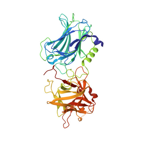
    The botulinum neurotoxins (BoNTs) are the most potent protein toxins for humans. There are seven serotypes of BoNTs (A-G) based on a lack of cross antiserum neutralization. BoNTs utilize gangliosides as components of the host receptors for binding and entry into neurons. Members of BoNT/C and BoNT/D serotypes include mosaic toxins that are organized in D/C and C/D toxins. One D/C mosaic toxin, BoNT/D-South Africa (BoNT/D-SA), was not fully neutralized by immunization with BoNT serotype C or D, which stimulated this study. Here the crystal structures of the receptor binding domains of BoNT/C, BoNT/D, and BoNT/D-SA are presented. Biochemical and cell binding studies show that BoNT/C and BoNT/D-SA possess unique mechanisms for ganglioside binding. These studies provide new information about how the BoNTs can enter host cells as well as a basis for understanding the immunological diversity of these neurotoxins.


  • Organizational Affiliation

    Department of Microbiology and of Molecular Genetics, Medical College of Wisconsin,Milwaukee, Wisconsin 53226-3548, USA.


Macromolecules
Find similar proteins by:  (by identity cutoff)  |  3D Structure
Entity ID: 1
MoleculeChains Sequence LengthOrganismDetailsImage
Botulinum neurotoxin type D415Clostridium botulinumMutation(s): 0 
Gene Names: botDClostridium phage d-16 phi
UniProt
Find proteins for P19321 (Clostridium botulinum D phage)
Explore P19321 
Go to UniProtKB:  P19321
Entity Groups  
Sequence Clusters30% Identity50% Identity70% Identity90% Identity95% Identity100% Identity
UniProt GroupP19321
Sequence Annotations
Expand
  • Reference Sequence
Experimental Data & Validation

Experimental Data

  • Method: X-RAY DIFFRACTION
  • Resolution: 2.00 Å
  • R-Value Free: 0.256 
  • R-Value Work: 0.222 
  • R-Value Observed: 0.222 
  • Space Group: P 21 21 21
Unit Cell:
Length ( Å )Angle ( ˚ )
a = 60.04α = 90
b = 94.15β = 90
c = 94.99γ = 90
Software Package:
Software NamePurpose
CrystalCleardata collection
MOLREPphasing
CNSrefinement
HKL-2000data reduction
HKL-2000data scaling

Structure Validation

View Full Validation Report


Entry History 

Deposition Data

Revision History  (Full details and data files)

  • Version 1.0: 2010-09-08
    Type: Initial release
  • Version 1.1: 2011-07-13
    Changes: Version format compliance
  • Version 1.2: 2015-01-28
    Changes: Structure summary
  • Version 1.3: 2023-09-06
    Changes: Data collection, Database references, Refinement description