3N7B

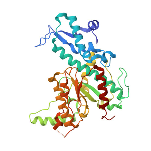
SgrAI bound to secondary site DNA and Ca(II)


Experimental Data Snapshot

  • Method: X-RAY DIFFRACTION
  • Resolution: 2.65 Å
  • R-Value Free: 0.248 
  • R-Value Work: 0.173 
  • R-Value Observed: 0.176 

wwPDB Validation   3D Report Full Report


This is version 1.3 of the entry. See complete history


Literature

New clues in the allosteric activation of DNA cleavage by SgrAI: structures of SgrAI bound to cleaved primary-site DNA and uncleaved secondary-site DNA.

Little, E.J.Dunten, P.W.Bitinaite, J.Horton, N.C.

(2011) Acta Crystallogr D Biol Crystallogr 67: 67-74

  • DOI: https://doi.org/10.1107/S0907444910047785
  • Primary Citation of Related Structures:  
    3MQY, 3N78, 3N7B

  • PubMed Abstract: 

    SgrAI is a type II restriction endonuclease that cuts an unusually long recognition sequence and exhibits allosteric self-activation with expansion of DNA-sequence specificity. The three-dimensional crystal structures of SgrAI bound to cleaved primary-site DNA and Mg²(+) and bound to secondary-site DNA with either Mg²(+) or Ca²(+) are presented. All three structures show a conformation of enzyme and DNA similar to the previously determined dimeric structure of SgrAI bound to uncleaved primary-site DNA and Ca²(+) [Dunten et al. (2008), Nucleic Acids Res. 36, 5405-5416], with the exception of the cleaved bond and a slight shifting of the DNA in the SgrAI/cleaved primary-site DNA/Mg²(+) structure. In addition, a new metal ion binding site is located in one of the two active sites in this structure, which is consistent with proposals for the existence of a metal-ion site near the 3'-O leaving group.


  • Organizational Affiliation

    Department of Chemistry and Biochemistry, University of Arizona, Tucson, 85721, USA.


Macromolecules

Find similar proteins by:  (by identity cutoff)  |  3D Structure
Entity ID: 1
MoleculeChains Sequence LengthOrganismDetailsImage
SgraIR restriction enzyme
A, B
338Streptomyces griseusMutation(s): 1 
Gene Names: sgraIR
EC: 3.1.21.4
UniProt
Find proteins for Q9F6L0 (Streptomyces griseus)
Explore Q9F6L0 
Go to UniProtKB:  Q9F6L0
Entity Groups  
Sequence Clusters30% Identity50% Identity70% Identity90% Identity95% Identity100% Identity
UniProt GroupQ9F6L0
Sequence Annotations
Expand
  • Reference Sequence

Find similar nucleic acids by:  Sequence   |   3D Structure  

Entity ID: 2
MoleculeChains LengthOrganismImage
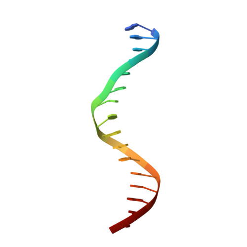
DNA (5'-D(*AP*GP*TP*CP*CP*AP*CP*CP*GP*GP*GP*GP*GP*AP*CP*T)-3')16N/A
Sequence Annotations
Expand
  • Reference Sequence

Find similar nucleic acids by:  Sequence   |   3D Structure  

Entity ID: 3
MoleculeChains LengthOrganismImage
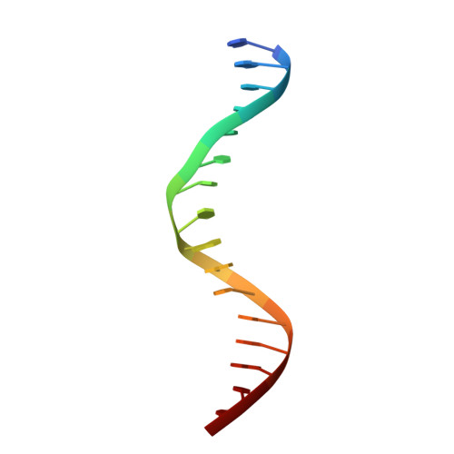
DNA (5'-D(*AP*GP*TP*CP*CP*CP*CP*CP*GP*GP*TP*GP*GP*AP*CP*T)-3')16N/A
Sequence Annotations
Expand
  • Reference Sequence
Experimental Data & Validation

Experimental Data

  • Method: X-RAY DIFFRACTION
  • Resolution: 2.65 Å
  • R-Value Free: 0.248 
  • R-Value Work: 0.173 
  • R-Value Observed: 0.176 
  • Space Group: P 21 21 2
Unit Cell:
Length ( Å )Angle ( ˚ )
a = 108.682α = 90
b = 133.662β = 90
c = 64.748γ = 90
Software Package:
Software NamePurpose
REFMACrefinement
SCALAdata scaling
PHENIXrefinement
PDB_EXTRACTdata extraction
Blu-Icedata collection
PHASERphasing
MOSFLMdata reduction

Structure Validation

View Full Validation Report


Entry History 

Deposition Data

Revision History  (Full details and data files)

  • Version 1.0: 2010-11-24
    Type: Initial release
  • Version 1.1: 2011-07-13
    Changes: Version format compliance
  • Version 1.2: 2019-07-17
    Changes: Data collection, Refinement description
  • Version 1.3: 2024-02-21
    Changes: Data collection, Database references, Derived calculations