


Crystal structure of PDE9A (E406A) mutation in complex with IBMX
Hou, J., Luo, H.-B., Chen, Y., Xu, J., Zhao, R., Zou, L.To be published.
Experimental Data Snapshot
Starting Model: experimental
View more details
Entity ID: 1 | |||||
|---|---|---|---|---|---|
| Molecule | Chains | Sequence Length | Organism | Details | Image |
| High affinity cGMP-specific 3',5'-cyclic phosphodiesterase 9A | 326 | Homo sapiens | Mutation(s): 1 Gene Names: PDE EC: 3.1.4.35 | 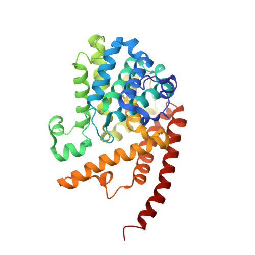 | |
UniProt & NIH Common Fund Data Resources | |||||
PHAROS: O76083 GTEx: ENSG00000160191 | |||||
Entity Groups | |||||
| Sequence Clusters | 30% Identity50% Identity70% Identity90% Identity95% Identity100% Identity | ||||
| UniProt Group | O76083 | ||||
Sequence AnnotationsExpand | |||||
Reference Sequence | |||||
| Ligands 4 Unique | |||||
|---|---|---|---|---|---|
| ID | Chains | Name / Formula / InChI Key | 2D Diagram | 3D Interactions | |
| IBM Download:Ideal Coordinates CCD File | C [auth A], H [auth B] | 3-ISOBUTYL-1-METHYLXANTHINE C10 H14 N4 O2 APIXJSLKIYYUKG-UHFFFAOYSA-N |  | ||
| ZN Download:Ideal Coordinates CCD File | D [auth A], F [auth B] | ZINC ION Zn PTFCDOFLOPIGGS-UHFFFAOYSA-N |  | ||
| CL Download:Ideal Coordinates CCD File | I [auth B] | CHLORIDE ION Cl VEXZGXHMUGYJMC-UHFFFAOYSA-M |  | ||
| MG Download:Ideal Coordinates CCD File | E [auth A], G [auth B] | MAGNESIUM ION Mg JLVVSXFLKOJNIY-UHFFFAOYSA-N |  | ||
| Length ( Å ) | Angle ( ˚ ) |
|---|---|
| a = 104.09 | α = 90 |
| b = 104.09 | β = 90 |
| c = 270.21 | γ = 90 |
| Software Name | Purpose |
|---|---|
| MAR345dtb | data collection |
| MOLREP | phasing |
| REFMAC | refinement |
| HKL-2000 | data reduction |
| HKL-2000 | data scaling |


