Probing the Impact of the echinT C-Terminal Domain on Structure and Catalysis.
Bardaweel, S., Pace, J., Chou, T.F., Cody, V., Wagner, C.R.(2010) J Mol Biology 404: 627-638
- PubMed: 20934431 Search on PubMed
- DOI: https://doi.org/10.1016/j.jmb.2010.09.066
- Primary Citation Related Structures:
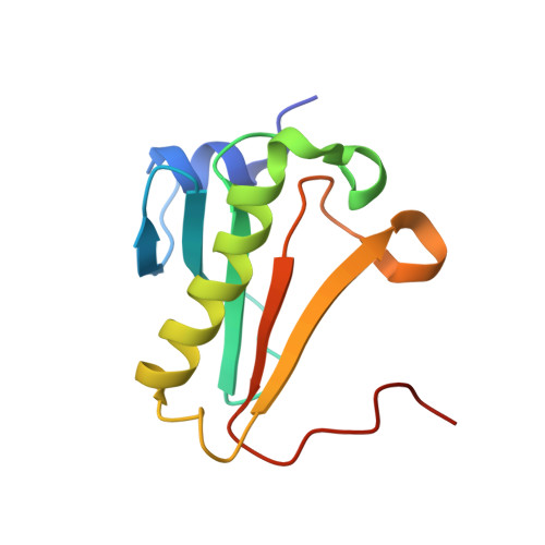
3N1S, 3N1T - PubMed Abstract:
Histidine triad nucleotide binding protein (Hint) is considered as the ancestor of the histidine triad protein superfamily and is highly conserved from bacteria to humans. Prokaryote genomes, including a wide array of both Gram-negative bacteria and Gram-positive bacteria, typically encode one Hint gene. The cellular function of Hint and the rationale for its evolutionary conservation in bacteria have remained a mystery. Despite its ubiquity and high sequence similarity to eukaryote Hint1 [Escherichia coli Hint (echinT) is 48% identical with human Hint1], prokaryote Hint has been reported in only a few studies. Here we report the first conformational information on the full-length N-terminal and C-terminal residues of Hint from the E. coli complex with GMP. Structural analysis of the echinT-GMP complex reveals that it crystallizes in the monoclinic space group P2(1) with four homodimers in the asymmetric unit. Analysis of electron density for both the N-terminal residues and the C-terminal residues of the echinT-GMP complex indicates that the loops in some monomers can adopt more than one conformation. The observation of conformational flexibility in terminal loop regions could explain the presence of multiple homodimers in the asymmetric unit of this structure. To explore the impact of the echinT C-terminus on protein structure and catalysis, we conducted a series of catalytic radiolabeling and kinetic experiments on the C-terminal deletion mutants of echinT. In this study, we show that sequential deletion of the C-terminus likely has no effect on homodimerization and a modest effect on the secondary structure of echinT. However, we observed a significant impact on the folding structure, as reflected by a significant lowering of the T(m) value. Kinetic analysis reveals that the C-terminal deletion mutants are within an order of magnitude less efficient in catalysis compared to wild type, while the overall kinetic mechanism that proceeds through a fast step, followed by a rate-limiting hydrolysis step, was conserved. Nevertheless, the ability of the C-terminal deletion mutants to hydrolyze lysyl-AMP generated by LysU was greatly impaired. Taken together, our results highlight the emerging role of the C-terminus in governing the catalytic function of Hints.
- Department of Medicinal Chemistry, University of Minnesota, 8-174 Weaver Densford Hall, 308 Harvard Street SE, Minneapolis, MN 55455, USA.
Organizational Affiliation: