3MYJ | pdb_00003myj

Human Class I MHC HLA-A2 in complex with the WT-1 (126-134) (R1Y) peptide variant.


Experimental Data Snapshot

  • Method: X-RAY DIFFRACTION
  • Resolution: 1.89 Å
  • R-Value Free: 
    0.230 (Depositor), 0.239 (DCC) 
  • R-Value Work: 
    0.185 (Depositor), 0.196 (DCC) 
  • R-Value Observed: 
    0.188 (Depositor) 

Starting Model: experimental
View more details

wwPDB Validation 3D Report Full Report

Validation slider image for 3MYJ

This is version 1.4 of the entry. See complete history

Literature

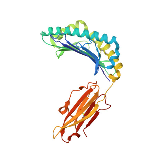
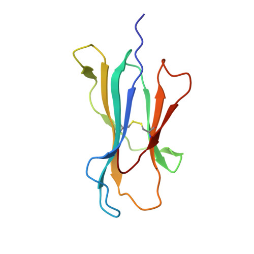
Structures of native and affinity-enhanced WT1 epitopes bound to HLA-A*0201: Implications for WT1-based cancer therapeutics.

Borbulevych, O.Y.Do, P.Baker, B.M.

(2010) Mol Immunol 47: 2519-2524

  • DOI: https://doi.org/10.1016/j.molimm.2010.06.005
  • Primary Citation Related Structures: 
    3HPJ, 3MYJ

  • PubMed Abstract: 

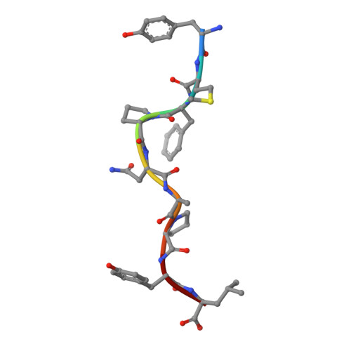
    Presentation of peptides by class I or class II major histocompatibility complex (MHC) molecules is required for the initiation and propagation of a T cell-mediated immune response. Peptides from the Wilms Tumor 1 transcription factor (WT1), upregulated in many hematopoetic and solid tumors, can be recognized by T cells and numerous efforts are underway to engineer WT1-based cancer vaccines. Here we determined the structures of the class I MHC molecule HLA-A*0201 bound to the native 126-134 epitope of the WT1 peptide and a recently described variant (R1Y) with improved MHC binding. The R1Y variant, a potential vaccine candidate, alters the positions of MHC charged side chains near the peptide N-terminus and significantly reduces the peptide/MHC electrostatic surface potential. These alterations indicate that the R1Y variant is an imperfect mimic of the native WT1 peptide, and suggest caution in its use as a therapeutic vaccine. Stability measurements revealed how the R1Y substitution enhances MHC binding affinity, and together with the structures suggest a strategy for engineering WT1 variants with improved MHC binding that retain the structural features of the native peptide/MHC complex.


  • Organizational Affiliation
    • Department of Chemistry and Biochemistry, University of Notre Dame, 251 Nieuwland Science Hall, Notre Dame, IN 46556, United States.

Macromolecule Content 

  • Total Structure Weight: 89.79 kDa 
  • Atom Count: 6,895 
  • Modeled Residue Count: 768 
  • Deposited Residue Count: 768 
  • Unique protein chains: 3

Macromolecules

Find similar proteins by:|  3D Structure
Entity ID: 1
MoleculeChains  Sequence LengthOrganismDetailsImage
HLA class I histocompatibility antigen, A-2 alpha chain
A, D
275Homo sapiensMutation(s): 0 
Gene Names: HLA-AHLAA
UniProt & NIH Common Fund Data Resources
Find proteins for P04439 (Homo sapiens)
Explore P04439 
Go to UniProtKB:  P04439
PHAROS:  P04439
GTEx:  ENSG00000206503 
Entity Groups
Sequence Clusters30% Identity50% Identity70% Identity90% Identity95% Identity100% Identity
UniProt GroupP04439
Sequence Annotations
Expand
Reference Sequence
Find similar proteins by:|  3D Structure
Entity ID: 2
MoleculeChains  Sequence LengthOrganismDetailsImage
Beta-2-microglobulin
B, E
100Homo sapiensMutation(s): 0 
Gene Names: B2MCDABP0092HDCMA22P
UniProt & NIH Common Fund Data Resources
Find proteins for P61769 (Homo sapiens)
Explore P61769 
Go to UniProtKB:  P61769
PHAROS:  P61769
GTEx:  ENSG00000166710 
Entity Groups
Sequence Clusters30% Identity50% Identity70% Identity90% Identity95% Identity100% Identity
UniProt GroupP61769
Sequence Annotations
Expand
Reference Sequence
Find similar proteins by:  Sequence   |   3D Structure  
Entity ID: 3
MoleculeChains  Sequence LengthOrganismDetailsImage
Wilms tumor protein
C, F
9Homo sapiensMutation(s): 1 
UniProt & NIH Common Fund Data Resources
Find proteins for P19544 (Homo sapiens)
Explore P19544 
Go to UniProtKB:  P19544
PHAROS:  P19544
GTEx:  ENSG00000184937 
Entity Groups
UniProt GroupP19544
Sequence Annotations
Expand
Reference Sequence

Small Molecules

Ligands 1 Unique
IDChains Name / Formula / InChI Key2D Diagram3D Interactions
GOL

Query on GOL


Download:Ideal Coordinates CCD File
G [auth A]GLYCEROL
C3 H8 O3
PEDCQBHIVMGVHV-UHFFFAOYSA-N

Experimental Data & Validation

Experimental Data

  • Method: X-RAY DIFFRACTION
  • Resolution: 1.89 Å
  • R-Value Free:  0.230 (Depositor), 0.239 (DCC) 
  • R-Value Work:  0.185 (Depositor), 0.196 (DCC) 
  • R-Value Observed: 0.188 (Depositor) 
Space Group: P 1
Unit Cell:
Length ( Å )Angle ( ˚ )
a = 50.202α = 81.91
b = 62.826β = 75.9
c = 74.755γ = 77.94
Software Package:
Software NamePurpose
DENZOdata reduction
SCALEPACKdata scaling
MOLREPphasing
REFMACrefinement
PDB_EXTRACTdata extraction
SBC-Collectdata collection
HKL-2000data reduction
HKL-2000data scaling

Structure Validation

View Full Validation Report


Entry History 

Deposition Data

Revision History  (Full details and data files)

  • Version 1.0: 2010-08-18
    Type: Initial release
  • Version 1.1: 2011-07-13
    Changes: Version format compliance
  • Version 1.2: 2017-11-08
    Changes: Refinement description
  • Version 1.3: 2023-09-06
    Changes: Data collection, Database references, Derived calculations, Refinement description
  • Version 1.4: 2024-11-06
    Changes: Structure summary