3MYA | pdb_00003mya

Crystal Structure of HP67 H41F - P61


Experimental Data Snapshot

  • Method: X-RAY DIFFRACTION
  • Resolution: 2.50 Å
  • R-Value Free: 
    0.277 (Depositor), 0.261 (DCC) 
  • R-Value Work: 
    0.215 (Depositor), 0.214 (DCC) 
  • R-Value Observed: 
    0.217 (Depositor) 

Starting Model: experimental
View more details

wwPDB Validation 3D Report Full Report

Validation slider image for 3MYA

This is version 1.1 of the entry. See complete history

Literature

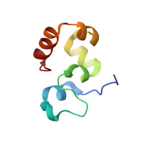
On unsatisfied hydrogen bonds in the N-terminal subdomain of villin headpiece.

Brown, J.W.Farelli, J.D.McKnight, C.J.

(2011) J Mol Biology 413: 543-547

  • DOI: https://doi.org/10.1016/j.jmb.2011.08.024
  • Primary Citation Related Structures: 
    3MYA, 3MYC

  • PubMed Abstract: 

    Villin headpiece is a small autonomously folding protein that has emerged as a model system for understanding the fundamental tenets governing protein folding. In this communication, we employ NMR and X-ray crystallography to characterize a point mutant, H41F, which retains actin-binding activity, is more thermostable but, interestingly, does not exhibit the partially folded intermediate observed of either wild-type or other similar point mutants.


  • Organizational Affiliation
    • Department of Physiology and Biophysics, Boston University School of Medicine, Boston, MA 02118, USA.

Macromolecule Content 

  • Total Structure Weight: 15.24 kDa 
  • Atom Count: 1,081 
  • Modeled Residue Count: 131 
  • Deposited Residue Count: 134 
  • Unique protein chains: 1

Macromolecules

Find similar proteins by:|  3D Structure
Entity ID: 1
MoleculeChains  Sequence LengthOrganismDetailsImage
Villin-1
A, B
67Gallus gallusMutation(s): 1 
Gene Names: VILVIL1
UniProt
Find proteins for P02640 (Gallus gallus)
Explore P02640 
Go to UniProtKB:  P02640
Entity Groups
Sequence Clusters30% Identity50% Identity70% Identity90% Identity95% Identity100% Identity
UniProt GroupP02640
Sequence Annotations
Expand
Reference Sequence

Experimental Data & Validation

Experimental Data

  • Method: X-RAY DIFFRACTION
  • Resolution: 2.50 Å
  • R-Value Free:  0.277 (Depositor), 0.261 (DCC) 
  • R-Value Work:  0.215 (Depositor), 0.214 (DCC) 
  • R-Value Observed: 0.217 (Depositor) 
Space Group: P 61
Unit Cell:
Length ( Å )Angle ( ˚ )
a = 65.409α = 90
b = 65.409β = 90
c = 68.283γ = 120
Software Package:
Software NamePurpose
CrystalCleardata collection
PHENIXmodel building
PHENIXrefinement
DENZOdata reduction
SCALEPACKdata scaling
PHENIXphasing

Structure Validation

View Full Validation Report


Entry History 

Deposition Data

Revision History  (Full details and data files)

  • Version 1.0: 2011-06-29
    Type: Initial release
  • Version 1.1: 2023-09-06
    Changes: Data collection, Database references, Refinement description