3MXN | pdb_00003mxn

Crystal structure of the RMI core complex


Experimental Data Snapshot

  • Method: X-RAY DIFFRACTION
  • Resolution: 1.55 Å
  • R-Value Free: 
    0.231 (Depositor), 0.234 (DCC) 
  • R-Value Work: 
    0.195 (Depositor), 0.200 (DCC) 
  • R-Value Observed: 
    0.197 (Depositor) 

wwPDB Validation 3D Report Full Report

Validation slider image for 3MXN

This is version 1.3 of the entry. See complete history

Literature

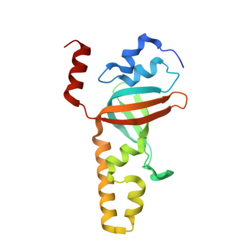
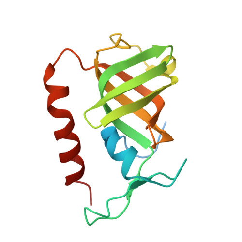
Structure and cellular roles of the RMI core complex from the bloom syndrome dissolvasome.

Hoadley, K.A.Xu, D.Xue, Y.Satyshur, K.A.Wang, W.Keck, J.L.

(2010) Structure 18: 1149-1158

  • DOI: https://doi.org/10.1016/j.str.2010.06.009
  • Primary Citation Related Structures: 
    3MXN

  • PubMed Abstract: 

    BLM, the protein product of the gene mutated in Bloom syndrome, is one of five human RecQ helicases. It functions to separate double Holliday junction DNA without genetic exchange as a component of the "dissolvasome," which also includes topoisomerase IIIα and the RMI (RecQ-mediated genome instability) subcomplex (RMI1 and RMI2). We describe the crystal structure of the RMI core complex, comprising RMI2 and the C-terminal OB domain of RMI1. The overall RMI core structure strongly resembles two-thirds of the trimerization core of the eukaryotic single-stranded DNA-binding protein, Replication Protein A. Immunoprecipitation experiments with RMI2 variants confirm key interactions that stabilize the RMI core interface. Disruption of this interface leads to a dramatic increase in cellular sister chromatid exchange events similar to that seen in BLM-deficient cells. The RMI core interface is therefore crucial for BLM dissolvasome assembly and may have additional cellular roles as a docking hub for other proteins.


  • Organizational Affiliation
    • Department of Biomolecular Chemistry, University of Wisconsin School of Medicine and Public Health, 550 Medical Science Center, 1300 University Avenue, Madison, WI 53706-1532, USA.

Macromolecule Content 

  • Total Structure Weight: 33.69 kDa 
  • Atom Count: 2,523 
  • Modeled Residue Count: 282 
  • Deposited Residue Count: 307 
  • Unique protein chains: 2

Macromolecules

Find similar proteins by:|  3D Structure
Entity ID: 1
MoleculeChains  Sequence LengthOrganismDetailsImage
RecQ-mediated genome instability protein 1157Homo sapiensMutation(s): 0 
Gene Names: RMI1C9orf76
UniProt & NIH Common Fund Data Resources
Find proteins for Q9H9A7 (Homo sapiens)
Explore Q9H9A7 
Go to UniProtKB:  Q9H9A7
PHAROS:  Q9H9A7
GTEx:  ENSG00000178966 
Entity Groups
Sequence Clusters30% Identity50% Identity70% Identity90% Identity95% Identity100% Identity
UniProt GroupQ9H9A7
Sequence Annotations
Expand
Reference Sequence
Find similar proteins by:|  3D Structure
Entity ID: 2
MoleculeChains  Sequence LengthOrganismDetailsImage
RecQ-mediated genome instability protein 2150Homo sapiensMutation(s): 0 
Gene Names: RMI2C16orf75
UniProt & NIH Common Fund Data Resources
Find proteins for Q96E14 (Homo sapiens)
Explore Q96E14 
Go to UniProtKB:  Q96E14
PHAROS:  Q96E14
GTEx:  ENSG00000175643 
Entity Groups
Sequence Clusters30% Identity50% Identity70% Identity90% Identity95% Identity100% Identity
UniProt GroupQ96E14
Sequence Annotations
Expand
Reference Sequence

Small Molecules

Ligands 1 Unique
IDChains Name / Formula / InChI Key2D Diagram3D Interactions
BEN

Query on BEN


Download:Ideal Coordinates CCD File
C [auth A]BENZAMIDINE
C7 H8 N2
PXXJHWLDUBFPOL-UHFFFAOYSA-N

Experimental Data & Validation

Experimental Data

  • Method: X-RAY DIFFRACTION
  • Resolution: 1.55 Å
  • R-Value Free:  0.231 (Depositor), 0.234 (DCC) 
  • R-Value Work:  0.195 (Depositor), 0.200 (DCC) 
  • R-Value Observed: 0.197 (Depositor) 
Space Group: P 21 21 21
Unit Cell:
Length ( Å )Angle ( ˚ )
a = 42.096α = 90
b = 42.223β = 90
c = 152.24γ = 90
Software Package:
Software NamePurpose
MAR345data collection
ARP/wARPmodel building
REFMACrefinement
HKL-2000data reduction
HKL-2000data scaling

Structure Validation

View Full Validation Report


Entry History 

Deposition Data

Revision History  (Full details and data files)

  • Version 1.0: 2010-09-15
    Type: Initial release
  • Version 1.1: 2011-07-13
    Changes: Version format compliance
  • Version 1.2: 2017-11-08
    Changes: Refinement description
  • Version 1.3: 2024-02-21
    Changes: Data collection, Database references, Derived calculations