3MVC

High resolution crystal structure of the heme domain of GLB-6 from C. elegans


Experimental Data Snapshot

  • Method: X-RAY DIFFRACTION
  • Resolution: 1.40 Å
  • R-Value Free: 0.205 
  • R-Value Work: 0.161 
  • R-Value Observed: 0.163 

wwPDB Validation   3D Report Full Report


This is version 1.2 of the entry. See complete history


Literature

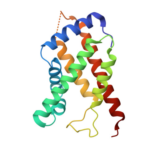
Structure and properties of a bis-histidyl ligated globin from Caenorhabditis elegans.

Yoon, J.Herzik, M.A.Winter, M.B.Tran, R.Olea, C.Marletta, M.A.

(2010) Biochemistry 49: 5662-5670

  • DOI: https://doi.org/10.1021/bi100710a
  • Primary Citation of Related Structures:  
    3MVC

  • PubMed Abstract: 

    Globins are heme-containing proteins that are best known for their roles in oxygen (O(2)) transport and storage. However, more diverse roles of globins in biology are being revealed, including gas and redox sensing. In the nematode Caenorhabditis elegans, 33 globin or globin-like genes were recently identified, some of which are known to be expressed in the sensory neurons of the worm and linked to O(2) sensing behavior. Here, we describe GLB-6, a novel globin-like protein expressed in the neurons of C. elegans. Recombinantly expressed full-length GLB-6 contains a heme site with spectral features that are similar to those of other bis-histidyl ligated globins, such as neuroglobin and cytoglobin. In contrast to these globins, however, ligands such as CO, NO, and CN(-) do not bind to the heme in GLB-6, demonstrating that the endogenous histidine ligands are likely very tightly coordinated. Additionally, GLB-6 exhibits rapid two-state autoxidation kinetics in the presence of physiological O(2) levels as well as a low redox potential (-193 +/- 2 mV). A high-resolution (1.40 A) crystal structure of the ferric form of the heme domain of GLB-6 confirms both the putative globin fold and bis-histidyl ligation and also demonstrates key structural features that can be correlated with the unusual ligand binding and redox properties exhibited by the full-length protein. Taken together, the biochemical properties of GLB-6 suggest that this neural protein would most likely serve as a physiological sensor for O(2) in C. elegans via redox signaling and/or electron transfer.


  • Organizational Affiliation

    California Institute for Quantitative Biosciences, University of California, Berkeley, California 94720, USA.


Macromolecules
Find similar proteins by:  (by identity cutoff)  |  3D Structure
Entity ID: 1
MoleculeChains Sequence LengthOrganismDetailsImage
Globin protein 6
A, B
161Caenorhabditis elegansMutation(s): 0 
Gene Names: C18C4.9glb-6
UniProt
Find proteins for Q18086 (Caenorhabditis elegans)
Explore Q18086 
Go to UniProtKB:  Q18086
Entity Groups  
Sequence Clusters30% Identity50% Identity70% Identity90% Identity95% Identity100% Identity
UniProt GroupQ18086
Sequence Annotations
Expand
  • Reference Sequence
Small Molecules
Ligands 3 Unique
IDChains Name / Formula / InChI Key2D Diagram3D Interactions
HEM
Query on HEM

Download Ideal Coordinates CCD File 
C [auth A],
M [auth B]
PROTOPORPHYRIN IX CONTAINING FE
C34 H32 Fe N4 O4
KABFMIBPWCXCRK-RGGAHWMASA-L
PR
Query on PR

Download Ideal Coordinates CCD File 
D [auth A],
E [auth A],
F [auth A],
N [auth B]
PRASEODYMIUM ION
Pr
WCWKKSOQLQEJTE-UHFFFAOYSA-N
NO3
Query on NO3

Download Ideal Coordinates CCD File 
G [auth A]
H [auth A]
I [auth A]
J [auth A]
K [auth A]
G [auth A],
H [auth A],
I [auth A],
J [auth A],
K [auth A],
L [auth A],
O [auth B],
P [auth B],
Q [auth B]
NITRATE ION
N O3
NHNBFGGVMKEFGY-UHFFFAOYSA-N
Experimental Data & Validation

Experimental Data

  • Method: X-RAY DIFFRACTION
  • Resolution: 1.40 Å
  • R-Value Free: 0.205 
  • R-Value Work: 0.161 
  • R-Value Observed: 0.163 
  • Space Group: P 1 21 1
Unit Cell:
Length ( Å )Angle ( ˚ )
a = 37.582α = 90
b = 76.347β = 91.76
c = 62.696γ = 90
Software Package:
Software NamePurpose
ADSCdata collection
SOLVEphasing
PHENIXrefinement
HKL-2000data reduction
SCALEPACKdata scaling

Structure Validation

View Full Validation Report


Entry History 

Deposition Data

Revision History  (Full details and data files)

  • Version 1.0: 2010-06-16
    Type: Initial release
  • Version 1.1: 2011-07-13
    Changes: Version format compliance
  • Version 1.2: 2024-02-21
    Changes: Data collection, Database references, Derived calculations