Structural insight into the mechanisms of enveloped virus tethering by tetherin.
Yang, H., Wang, J., Jia, X., McNatt, M.W., Zang, T., Pan, B., Meng, W., Wang, H.W., Bieniasz, P.D., Xiong, Y.(2010) Proc Natl Acad Sci U S A 107: 18428-18432
- PubMed: 20940320 Search on PubMedSearch on PubMed Central
- DOI: https://doi.org/10.1073/pnas.1011485107
- Primary Citation Related Structures:
3MQ7, 3MQ9, 3MQB, 3MQC - PubMed Abstract:
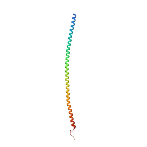
Tetherin/BST2 is a type-II membrane protein that inhibits the release of a range of enveloped viruses, including HIV-1. Here we report three crystal structures of human tetherin, including the full-length ectodomain, a triple cysteine mutant and an ectodomain truncation. These structures show that tetherin forms a continuous alpha helix encompassing almost the entire ectodomain. Tetherin helices dimerize into parallel coiled coils via interactions throughout the C-terminal portion of the ectodomain. A comparison of the multiple structures of the tetherin dimer reveals inherent constrained flexibility at two hinges positioned at residues A88 and G109. In the crystals, two tetherin ectodomain dimers associate into a tetramer by forming an antiparallel four-helix bundle at their N termini. However, mutagenesis studies suggest that the tetrametric form of tetherin, although potentially contributing to, is not essential for its antiviral activity. Nonetheless, the structural and chemical properties of the N terminus of the ectodomain are important for optimal tethering function. This study provides detailed insight into the mechanisms by which this broad-spectrum antiviral restriction factor can function.
- Department of Molecular Biophysics and Biochemistry, Yale University, New Haven, CT 06520-8114, USA.
Organizational Affiliation: