3MMG

Crystal structure of tobacco vein mottling virus protease


Experimental Data Snapshot

  • Method: X-RAY DIFFRACTION
  • Resolution: 1.70 Å
  • R-Value Free: 0.210 
  • R-Value Work: 0.175 
  • R-Value Observed: 0.176 

wwPDB Validation   3D Report Full Report


This is version 1.2 of the entry. See complete history


Literature

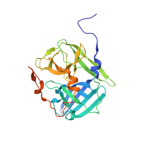
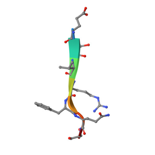
Structural determinants of tobacco vein mottling virus protease substrate specificity.

Sun, P.Austin, B.P.Tozser, J.Waugh, D.S.

(2010) Protein Sci 19: 2240-2251

  • DOI: https://doi.org/10.1002/pro.506
  • Primary Citation of Related Structures:  
    3MMG

  • PubMed Abstract: 

    Tobacco vein mottling virus (TVMV) is a member of the Potyviridae, one of the largest families of plant viruses. The TVMV genome is translated into a single large polyprotein that is subsequently processed by three virally encoded proteases. Seven of the nine cleavage events are carried out by the NIa protease. Its homolog from the tobacco etch virus (TEV) is a widely used reagent for the removal of affinity tags from recombinant proteins. Although TVMV protease is a close relative of TEV protease, they exhibit distinct sequence specificities. We report here the crystal structure of a catalytically inactive mutant TVMV protease (K65A/K67A/C151A) in complex with a canonical peptide substrate (Ac-RETVRFQSD) at 1.7-Å resolution. As observed in several crystal structures of TEV protease, the C-terminus (∼20 residues) of TVMV protease is disordered. Unexpectedly, although deleting the disordered residues from TEV protease reduces its catalytic activity by ∼10-fold, an analogous truncation mutant of TVMV protease is significantly more active. Comparison of the structures of TEV and TVMV protease in complex with their respective canonical substrate peptides reveals that the S3 and S4 pockets are mainly responsible for the differing substrate specificities. The structure of TVMV protease suggests that it is less tolerant of variation at the P1' position than TEV protease. This conjecture was confirmed experimentally by determining kinetic parameters k(cat) and K(m) for a series of oligopeptide substrates. Also, as predicted by the cocrystal structure, we confirm that substitutions in the P6 position are more readily tolerated by TVMV than TEV protease.


  • Organizational Affiliation

    Macromolecular Crystallography Laboratory, Center for Cancer Research, National Cancer Institute at Frederick, Frederick, Maryland, USA.


Macromolecules
Find similar proteins by:  (by identity cutoff)  |  3D Structure
Entity ID: 1
MoleculeChains Sequence LengthOrganismDetailsImage
Nuclear inclusion protein A
A, B
241Tobacco vein mottling virusMutation(s): 3 
Gene Names: protease
UniProt
Find proteins for P09814 (Tobacco vein mottling virus)
Explore P09814 
Go to UniProtKB:  P09814
Entity Groups  
Sequence Clusters30% Identity50% Identity70% Identity90% Identity95% Identity100% Identity
UniProt GroupP09814
Sequence Annotations
Expand
  • Reference Sequence

Find similar proteins by:  Sequence   |   3D Structure  

Entity ID: 2
MoleculeChains Sequence LengthOrganismDetailsImage
Nuclear inclusion protein B fragment
C, D
8Tobacco vein mottling virusMutation(s): 0 
UniProt
Find proteins for P09814 (Tobacco vein mottling virus)
Explore P09814 
Go to UniProtKB:  P09814
Entity Groups  
UniProt GroupP09814
Sequence Annotations
Expand
  • Reference Sequence
Small Molecules
Ligands 1 Unique
IDChains Name / Formula / InChI Key2D Diagram3D Interactions
FMT
Query on FMT

Download Ideal Coordinates CCD File 
E [auth A]FORMIC ACID
C H2 O2
BDAGIHXWWSANSR-UHFFFAOYSA-N
Experimental Data & Validation

Experimental Data

  • Method: X-RAY DIFFRACTION
  • Resolution: 1.70 Å
  • R-Value Free: 0.210 
  • R-Value Work: 0.175 
  • R-Value Observed: 0.176 
  • Space Group: P 21 21 21
Unit Cell:
Length ( Å )Angle ( ˚ )
a = 76.476α = 90
b = 77.563β = 90
c = 78.463γ = 90
Software Package:
Software NamePurpose
MAR345dtbdata collection
MOLREPphasing
REFMACrefinement
HKL-2000data reduction
HKL-2000data scaling

Structure Validation

View Full Validation Report


Entry History 

Deposition Data

Revision History  (Full details and data files)

  • Version 1.0: 2010-10-20
    Type: Initial release
  • Version 1.1: 2011-07-13
    Changes: Version format compliance
  • Version 1.2: 2023-09-06
    Changes: Data collection, Database references, Derived calculations, Refinement description