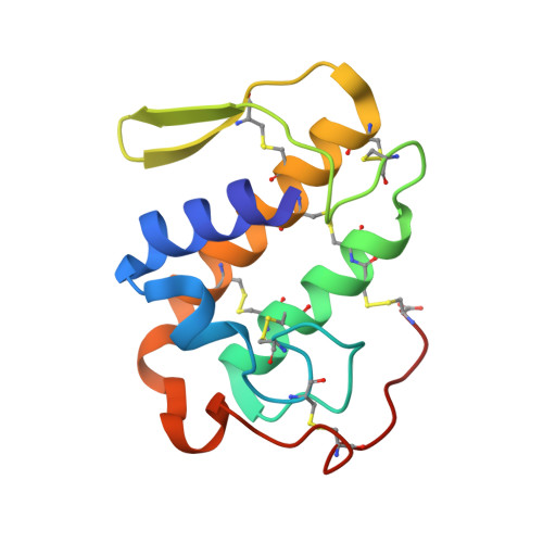
Crystal structure of Bn IV in complex with myristic acid: a Lys49 myotoxic phospholipase A2 from Bothrops neuwiedi venom.
Delatorre, P., Rocha, B.A., Santi-Gadelha, T., Gadelha, C.A., Toyama, M.H., Cavada, B.S.(2011) Biochimie 93: 513-518
- PubMed: 21108987 Search on PubMed
- DOI: https://doi.org/10.1016/j.biochi.2010.11.003
- Primary Citation Related Structures:
3MLM - PubMed Abstract:
The LYS49-PLA₂s myotoxins have attracted attention as models for the induction of myonecrosis by a catalytically independent mechanism of action. Structural studies and biological activities have demonstrated that the myotoxic activity of LYS49-PLA₂ is independent of the catalytic activity site. The myotoxic effect is conventionally thought to be to due to the C-terminal region 111-121, which plays an effective role in membrane damage. In the present study, Bn IV LYS49-PLA₂ was isolated from Bothrops neuwiedi snake venom in complex with myristic acid (CH₃(CH₂)₁₂COOH) and its overall structure was refined at 2.2 Å resolution. The Bn IV crystals belong to monoclinic space group P2₁ and contain a dimer in the asymmetric unit. The unit cell parameters are a = 38.8, b = 70.4, c = 44.0 Å. The biological assembly is a "conventional dimer" and the results confirm that dimer formation is not relevant to the myotoxic activity. Electron density map analysis of the Bn IV structure shows clearly the presence of myristic acid in catalytic site. The relevant structural features for myotoxic activity are located in the C-terminal region and the Bn IV C-terminal residues NKKYRY are a probable heparin binding domain. These findings indicate that the mechanism of interaction between Bn IV and muscle cell membranes is through some kind of cell signal transduction mediated by heparin complexes.
- Departamento de Biologia Molecular, Universidade Federal da Paraíba, João Pessoa, Paraíba, Brazil. pldelatorre@gmail.com
Organizational Affiliation: