3MKN

Crystal structure of the E. coli pyrimidine nucleosidase YeiK bound to a competitive inhibitor


Experimental Data Snapshot

  • Method: X-RAY DIFFRACTION
  • Resolution: 2.00 Å
  • R-Value Free: 0.196 
  • R-Value Work: 0.174 
  • R-Value Observed: 0.175 

wwPDB Validation   3D Report Full Report


Ligand Structure Quality Assessment 


This is version 1.2 of the entry. See complete history


Literature

Energy Landscapes Associated with Macromolecular Conformational Changes from Endpoint Structures

Fornili, A.Giabbai, B.Garau, G.Degano, M.

(2010) J Am Chem Soc 132: 17570-17577

  • DOI: https://doi.org/10.1021/ja107640u
  • Primary Citation of Related Structures:  
    3MKM, 3MKN

  • PubMed Abstract: 

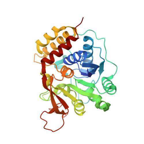
    Conformational changes modulate macromolecular function by promoting the specific binding of ligands (such as in antigen recognition) or the stabilization of transition states in enzymatic reactions. However, quantitative characterization of the energetics underlying dynamic structural interconversions is still challenging and lacks a unified method. Here, we introduce a novel in silico approach based on the combined use of essential dynamics sampling and nonequilibrium free-energy calculations to obtain quantitative data on conformational energy landscapes. This technique allows the unbiased investigation of highly complex rearrangements, and does not require the crucial definition of user-defined collective variables. We show that free-energy values derived from profiles connecting the unliganded and ligand-bound X-ray structures of a bacterial nucleoside hydrolase match the experimental binding constant. This approach also provides first evidence for a rate-limiting character of the conformational transition in this enzyme, and an unexpected role of the protonation state of a single residue in regulating substrate binding and product release.


  • Organizational Affiliation

    Biocrystallography Unit, Division of Immunology, Transplantation, and Infectious Diseases, Scientific Institute San Raffaele, Via Olgettina 58, 20132 Milan, Italy. arianna.fornili@kcl.ac.uk


Macromolecules
Find similar proteins by:  (by identity cutoff)  |  3D Structure
Entity ID: 1
MoleculeChains Sequence LengthOrganismDetailsImage
Putative uncharacterized protein YeiK
A, B, C, D
316Escherichia coli K-12Mutation(s): 0 
Gene Names: YeiK
EC: 3.2.2.8
UniProt
Find proteins for C3T3U2 (Escherichia coli)
Explore C3T3U2 
Go to UniProtKB:  C3T3U2
Entity Groups  
Sequence Clusters30% Identity50% Identity70% Identity90% Identity95% Identity100% Identity
UniProt GroupC3T3U2
Sequence Annotations
Expand
  • Reference Sequence
Small Molecules
Binding Affinity Annotations 
IDSourceBinding Affinity
DNB Binding MOAD:  3MKN Ki: 7.60e+4 (nM) from 1 assay(s)
PDBBind:  3MKN Ki: 7.60e+4 (nM) from 1 assay(s)
Experimental Data & Validation

Experimental Data

  • Method: X-RAY DIFFRACTION
  • Resolution: 2.00 Å
  • R-Value Free: 0.196 
  • R-Value Work: 0.174 
  • R-Value Observed: 0.175 
  • Space Group: P 1 21 1
Unit Cell:
Length ( Å )Angle ( ˚ )
a = 84.851α = 90
b = 86.446β = 98.86
c = 97.817γ = 90
Software Package:
Software NamePurpose
DNAdata collection
MOLREPphasing
REFMACrefinement
MOSFLMdata reduction
SCALAdata scaling

Structure Validation

View Full Validation Report


Ligand Structure Quality Assessment 


Entry History 

Deposition Data

Revision History  (Full details and data files)

  • Version 1.0: 2010-12-01
    Type: Initial release
  • Version 1.1: 2011-07-13
    Changes: Version format compliance
  • Version 1.2: 2023-11-01
    Changes: Data collection, Database references, Derived calculations, Refinement description