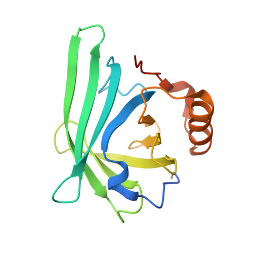
Structural and biochemical analyses reveal a monomeric state of the bacterial lipocalin Blc.
Schiefner, A., Chatwell, L., Breustedt, D.A., Skerra, A.(2010) Acta Crystallogr D Biol Crystallogr 66: 1308-1315
- PubMed: 21123871 Search on PubMed
- DOI: https://doi.org/10.1107/S0907444910039375
- Primary Citation Related Structures:
3MBT - PubMed Abstract:
The first bacterial member of the lipocalin protein family, Blc, was identified in Escherichia coli as an outer membrane lipoprotein that is expressed under conditions of environmental stress. Previous crystallographic studies in space group P2₁2₁2₁ with two molecules per asymmetric unit, supported by static light-scattering experiments in solution, indicated that Blc may form a functional homodimer with lysophospholipid binding activity. Here, a new crystal structure of recombinant Blc in space group I4₁22 with one molecule in the asymmetric unit is described. The crystal packing differs considerably from that observed previously, which was determined using an N-terminally extended version of Blc dubbed `Blc-X'. In particular, the characteristic large interface region that was previously described as being responsible for stable dimer formation is absent in the I4₁22 crystal structure. Thus, the dimerization behaviour of Blc-X was most likely to be caused by the additional N-terminal peptide segment resulting from the cloning strategy employed. In contrast, we used a native-like N-terminus for Blc with just the lipid-anchored first Cys residue replaced by Ala. The fully monomeric status of this recombinant version of Blc in solution was confirmed by size-exclusion chromatography as well as analytical ultracentrifugation. Consequently, these data shed new light on the previously postulated lipid-binding mechanism and biological role of Blc. Beyond this, our findings illustrate that cloning artefacts, which frequently result from recombinant protein production for structural studies, must be considered with special caution when interpreting oligomerization and/or conformational effects.
- Munich Center for Integrated Protein Science (CIPS-M) and Lehrstuhl für Biologische Chemie, Technische Universität München, Freising-Weihenstephan, Germany.
Organizational Affiliation: