


Crystal structure of a putative phosphomethylpyrimidine kinase (BT_4458) from BACTEROIDES THETAIOTAOMICRON VPI-5482 at 2.10 A resolution (rhombohedral form)
Joint Center for Structural Genomics (JCSG)To be published.
Experimental Data Snapshot
wwPDB Validation 3D Report Full Report

Entity ID: 1 | |||||
|---|---|---|---|---|---|
| Molecule | Chains | Sequence Length | Organism | Details | Image |
| Putative phosphomethylpyrimidine kinase | 291 | Bacteroides thetaiotaomicron VPI-5482 | Mutation(s): 0 Gene Names: BT_4458 EC: 2.7.1.35 | 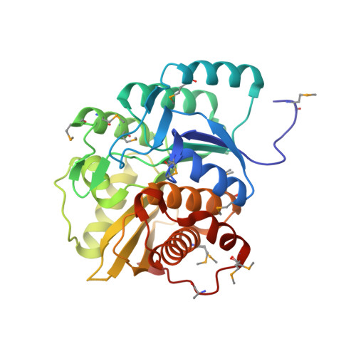 | |
UniProt | |||||
Entity Groups | |||||
| Sequence Clusters | 30% Identity50% Identity70% Identity90% Identity95% Identity100% Identity | ||||
| UniProt Group | Q89ZB9 | ||||
Sequence AnnotationsExpand | |||||
Reference Sequence | |||||
| Ligands 6 Unique | |||||
|---|---|---|---|---|---|
| ID | Chains | Name / Formula / InChI Key | 2D Diagram | 3D Interactions | |
| MPD Download:Ideal Coordinates CCD File | I [auth A], K [auth A] | (4S)-2-METHYL-2,4-PENTANEDIOL C6 H14 O2 SVTBMSDMJJWYQN-YFKPBYRVSA-N |  | ||
| MRD Download:Ideal Coordinates CCD File | J [auth A], L [auth A] | (4R)-2-METHYLPENTANE-2,4-DIOL C6 H14 O2 SVTBMSDMJJWYQN-RXMQYKEDSA-N |  | ||
| SO4 Download:Ideal Coordinates CCD File | F [auth A], G [auth A] | SULFATE ION O4 S QAOWNCQODCNURD-UHFFFAOYSA-L |  | ||
| IMD Download:Ideal Coordinates CCD File | H [auth A] | IMIDAZOLE C3 H5 N2 RAXXELZNTBOGNW-UHFFFAOYSA-O |  | ||
| ZN Download:Ideal Coordinates CCD File | B [auth A], C [auth A], D [auth A] | ZINC ION Zn PTFCDOFLOPIGGS-UHFFFAOYSA-N |  | ||
| CL Download:Ideal Coordinates CCD File | E [auth A] | CHLORIDE ION Cl VEXZGXHMUGYJMC-UHFFFAOYSA-M |  | ||
| Modified Residues 1 Unique | |||||
|---|---|---|---|---|---|
| ID | Chains | Type | Formula | 2D Diagram | Parent |
| MSE Query on MSE | A | L-PEPTIDE LINKING | C5 H11 N O2 Se |  | MET |
| Length ( Å ) | Angle ( ˚ ) |
|---|---|
| a = 161.627 | α = 90 |
| b = 161.627 | β = 90 |
| c = 76.138 | γ = 120 |
| Software Name | Purpose |
|---|---|
| REFMAC | refinement |
| PHENIX | refinement |
| SHELX | phasing |
| MolProbity | model building |
| SCALA | data scaling |
| PDB_EXTRACT | data extraction |
| MOSFLM | data reduction |
| autoSHARP | phasing |
| SHELXD | phasing |


