


Crystal Structure of Transketolase Complexed with Thiamine Diphosphate from Bacillus anthracis
Maltseva, N., Kim, Y., Kwon, K., Anderson, W.F., Joachimiak, A.To be published.
Experimental Data Snapshot
Entity ID: 1 | |||||
|---|---|---|---|---|---|
| Molecule | Chains | Sequence Length | Organism | Details | Image |
| Transketolase | 690 | Bacillus anthracis str. 'Ames Ancestor | Mutation(s): 0 Gene Names: BAS3470, BA_3744, GBAA3744, GBAA_3744, tkt-2, tkt2 EC: 2.2.1.1 | 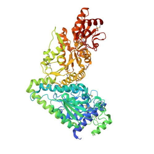 | |
UniProt | |||||
Entity Groups | |||||
| Sequence Clusters | 30% Identity50% Identity70% Identity90% Identity95% Identity100% Identity | ||||
| UniProt Group | A0A6L7H165 | ||||
Sequence AnnotationsExpand | |||||
Reference Sequence | |||||
| Ligands 10 Unique | |||||
|---|---|---|---|---|---|
| ID | Chains | Name / Formula / InChI Key | 2D Diagram | 3D Interactions | |
| TPP Download:Ideal Coordinates CCD File | AA [auth B], C [auth A] | THIAMINE DIPHOSPHATE C12 H19 N4 O7 P2 S AYEKOFBPNLCAJY-UHFFFAOYSA-O |  | ||
| BTB Download:Ideal Coordinates CCD File | CA [auth B] | 2-[BIS-(2-HYDROXY-ETHYL)-AMINO]-2-HYDROXYMETHYL-PROPANE-1,3-DIOL C8 H19 N O5 OWMVSZAMULFTJU-UHFFFAOYSA-N |  | ||
| PG5 Download:Ideal Coordinates CCD File | N [auth A] | 1-METHOXY-2-[2-(2-METHOXY-ETHOXY]-ETHANE C8 H18 O4 YFNKIDBQEZZDLK-UHFFFAOYSA-N |  | ||
| TRS Download:Ideal Coordinates CCD File | P [auth A] | 2-AMINO-2-HYDROXYMETHYL-PROPANE-1,3-DIOL C4 H12 N O3 LENZDBCJOHFCAS-UHFFFAOYSA-O |  | ||
| PEG Download:Ideal Coordinates CCD File | O [auth A], S [auth A] | DI(HYDROXYETHYL)ETHER C4 H10 O3 MTHSVFCYNBDYFN-UHFFFAOYSA-N |  | ||
| SO4 Download:Ideal Coordinates CCD File | BA [auth B] D [auth A] H [auth A] HA [auth B] I [auth A] | SULFATE ION O4 S QAOWNCQODCNURD-UHFFFAOYSA-L |  | ||
| GOL Download:Ideal Coordinates CCD File | EA [auth B] F [auth A] G [auth A] JA [auth B] LA [auth B] | GLYCEROL C3 H8 O3 PEDCQBHIVMGVHV-UHFFFAOYSA-N |  | ||
| ACY Download:Ideal Coordinates CCD File | M [auth A] | ACETIC ACID C2 H4 O2 QTBSBXVTEAMEQO-UHFFFAOYSA-N |  | ||
| FMT Download:Ideal Coordinates CCD File | DA [auth B] E [auth A] FA [auth B] GA [auth B] IA [auth B] | FORMIC ACID C H2 O2 BDAGIHXWWSANSR-UHFFFAOYSA-N |  | ||
| MG Download:Ideal Coordinates CCD File | R [auth A] | MAGNESIUM ION Mg JLVVSXFLKOJNIY-UHFFFAOYSA-N |  | ||
| Modified Residues 1 Unique | |||||
|---|---|---|---|---|---|
| ID | Chains | Type | Formula | 2D Diagram | Parent |
| MSE Query on MSE | A, B | L-PEPTIDE LINKING | C5 H11 N O2 Se |  | MET |
| Length ( Å ) | Angle ( ˚ ) |
|---|---|
| a = 83.333 | α = 90 |
| b = 132.087 | β = 90 |
| c = 137.319 | γ = 90 |
| Software Name | Purpose |
|---|---|
| SBC-Collect | data collection |
| HKL-3000 | data collection |
| HKL-3000 | phasing |
| SHELX | model building |
| MLPHARE | phasing |
| DM | model building |
| SOLVE | phasing |
| RESOLVE | model building |
| PHENIX | refinement |
| HKL-3000 | data reduction |
| HKL-3000 | data scaling |
| SHELX | phasing |
| DM | phasing |
| RESOLVE | phasing |


