3M3N | pdb_00003m3n

Structure of a Longitudinal Actin Dimer Assembled by Tandem W Domains


Experimental Data Snapshot

  • Method: X-RAY DIFFRACTION
  • Resolution: 7.00 Å
  • R-Value Work: 
    0.328 (DCC) 

Starting Model: experimental
View more details

wwPDB Validation 3D Report Full Report

Validation slider image for 3M3N

Ligand Structure Quality Assessment 


This is version 1.4 of the entry. See complete history

Literature

Structure of a longitudinal actin dimer assembled by tandem w domains: implications for actin filament nucleation.

Rebowski, G.Namgoong, S.Boczkowska, M.Leavis, P.C.Navaza, J.Dominguez, R.

(2010) J Mol Biology 403: 11-23

  • DOI: https://doi.org/10.1016/j.jmb.2010.08.040
  • Primary Citation Related Structures: 
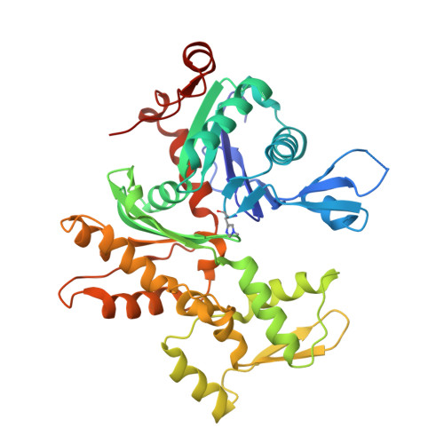
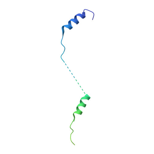
    3M1F, 3M3N

  • PubMed Abstract: 

    Actin filament nucleators initiate polymerization in cells in a regulated manner. A common architecture among these molecules consists of tandem WASP homology 2 domains (W domains) that recruit three to four actin subunits to form a polymerization nucleus. We describe a low-resolution crystal structure of an actin dimer assembled by tandem W domains, where the first W domain is cross-linked to Cys374 of the actin subunit bound to it, whereas the last W domain is followed by the C-terminal pointed end-capping helix of thymosin β4. While the arrangement of actin subunits in the dimer resembles that of a long-pitch helix of the actin filament, important differences are observed. These differences result from steric hindrance of the W domain with intersubunit contacts in the actin filament. We also determined the structure of the first W domain of Vibrio parahaemolyticus VopL cross-linked to actin Cys374 and show it to be nearly identical with non-cross-linked W-Actin structures. This result validates the use of cross-linking as a tool for the study of actin nucleation complexes, whose natural tendency to polymerize interferes with most structural methods. Combined with a biochemical analysis of nucleation, the structures may explain why nucleators based on tandem W domains with short inter-W linkers have relatively weak activity, cannot stay bound to filaments after nucleation, and are unlikely to influence filament elongation. The findings may also explain why nucleation-promoting factors of the Arp2/3 complex, which are related to tandem-W-domain nucleators, are ejected from branch junctions after nucleation. We finally show that the simple addition of the C-terminal pointed end-capping helix of thymosin β4 to tandem W domains can change their activity from actin filament nucleation to monomer sequestration.


  • Organizational Affiliation
    • Department of Physiology, University of Pennsylvania School of Medicine, 3700 Hamilton Walk, Philadelphia, PA 19104-6085, USA.

Macromolecule Content 

  • Total Structure Weight: 95.94 kDa 
  • Atom Count: 6,032 
  • Modeled Residue Count: 763 
  • Deposited Residue Count: 851 
  • Unique protein chains: 2

Macromolecules

Find similar proteins by:|  3D Structure
Entity ID: 1
MoleculeChains  Sequence LengthOrganismDetailsImage
Actin, alpha skeletal muscle
A, B
375Oryctolagus cuniculusMutation(s): 0 
EC: 3.6.4
UniProt
Find proteins for P68135 (Oryctolagus cuniculus)
Explore P68135 
Go to UniProtKB:  P68135
Entity Groups
Sequence Clusters30% Identity50% Identity70% Identity90% Identity95% Identity100% Identity
UniProt GroupP68135
Sequence Annotations
Expand
Reference Sequence
Find similar proteins by:|  3D Structure
Entity ID: 2
MoleculeChains  Sequence LengthOrganismDetailsImage
Neural Wiskott-Aldrich syndrome proteinC [auth W]101Mus musculusMutation(s): 3 
Gene Names: Wasl
UniProt & NIH Common Fund Data Resources
Find proteins for P20065 (Mus musculus)
Explore P20065 
Go to UniProtKB:  P20065
IMPC:  MGI:99510
Find proteins for Q91YD9 (Mus musculus)
Explore Q91YD9 
Go to UniProtKB:  Q91YD9
Entity Groups
Sequence Clusters30% Identity50% Identity70% Identity90% Identity95% Identity100% Identity
UniProt GroupsQ91YD9P20065
Sequence Annotations
Expand
Reference Sequence

Experimental Data & Validation

Experimental Data

  • Method: X-RAY DIFFRACTION
  • Resolution: 7.00 Å
  • R-Value Work:  0.328 (DCC) 
Space Group: P 65 2 2
Unit Cell:
Length ( Å )Angle ( ˚ )
a = 100.743α = 90
b = 100.743β = 90
c = 458.839γ = 120
Software Package:
Software NamePurpose
HKL-2000data collection
AMoREphasing
PHENIXmodel building
HKL-2000data reduction
HKL-2000data scaling
PHENIXphasing

Structure Validation

View Full Validation Report


Ligand Structure Quality Assessment 


Entry History 

Deposition Data

Revision History  (Full details and data files)

  • Version 1.0: 2010-07-28
    Type: Initial release
  • Version 1.1: 2011-07-13
    Changes: Version format compliance
  • Version 1.2: 2017-07-26
    Changes: Refinement description, Source and taxonomy
  • Version 1.3: 2021-10-06
    Changes: Database references, Derived calculations
  • Version 1.4: 2023-09-06
    Changes: Data collection, Refinement description