3M3L

PEPA bound to the ligand binding domain of GluA2 (flop form)


Experimental Data Snapshot

  • Method: X-RAY DIFFRACTION
  • Resolution: 1.85 Å
  • R-Value Free: 0.242 
  • R-Value Work: 0.187 
  • R-Value Observed: 0.189 

wwPDB Validation   3D Report Full Report


Ligand Structure Quality Assessment 


This is version 1.3 of the entry. See complete history


Literature

Molecular mechanism of flop selectivity and subsite recognition for an AMPA receptor allosteric modulator: structures of GluA2 and GluA3 in complexes with PEPA.

Ahmed, A.H.Ptak, C.P.Oswald, R.E.

(2010) Biochemistry 49: 2843-2850

  • DOI: https://doi.org/10.1021/bi1000678
  • Primary Citation of Related Structures:  
    3M3F, 3M3K, 3M3L

  • PubMed Abstract: 

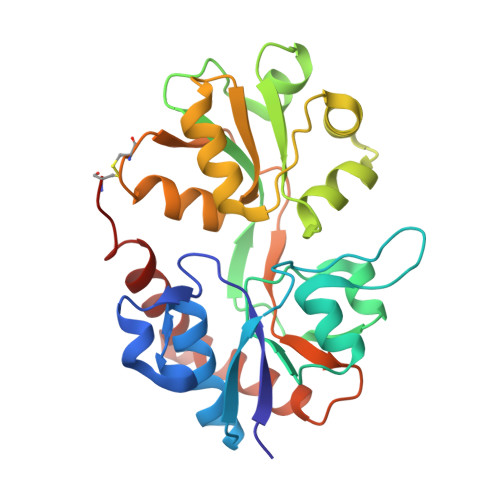
    Glutamate receptors are important potential drug targets for cognitive enhancement and the treatment of schizophrenia in part because they are the most prevalent excitatory neurotransmitter receptors in the vertebrate central nervous system. One approach to the application of therapeutic agents to the AMPA subtype of glutamate receptors is the use of allosteric modulators, which promote dimerization by binding to a dimer interface thereby reducing the degree of desensitization and deactivation. AMPA receptors exist in two alternatively spliced variants (flip and flop) that differ in desensitization and receptor activation profiles. Most of the structural information about modulators of the AMPA receptor targets the flip subtype. We report here the crystal structure of the flop-selective allosteric modulator, PEPA, bound to the binding domains of the GluA2 and GluA3 flop isoforms of AMPA receptors. Specific hydrogen bonding patterns can explain the preference for the flop isoform. This includes a bidentate hydrogen bonding pattern between PEPA and N754 of the flop isoforms of GluA2 and GluA3 (the corresponding position in the flip isoform is S754). Comparison with other allosteric modulators provides a framework for the development of new allosteric modulators with preferences for either the flip or flop isoforms. In addition to interactions with N/S754, specific interactions of the sulfonamide with conserved residues in the binding site are characteristics of a number of allosteric modulators. These, in combination with variable interactions with five subsites on the binding surface, lead to different stoichiometries, orientations within the binding pockets, and functional outcomes.


  • Organizational Affiliation

    Department of Molecular Medicine, Cornell University, Ithaca, New York 14853, USA.


Macromolecules
Find similar proteins by:  (by identity cutoff)  |  3D Structure
Entity ID: 1
MoleculeChains Sequence LengthOrganismDetailsImage
Glutamate receptor 2A,
B [auth D],
C [auth G]
258Rattus norvegicusMutation(s): 0 
Gene Names: Gria2Glur2
UniProt
Find proteins for P19491 (Rattus norvegicus)
Explore P19491 
Go to UniProtKB:  P19491
Entity Groups  
Sequence Clusters30% Identity50% Identity70% Identity90% Identity95% Identity100% Identity
UniProt GroupP19491
Sequence Annotations
Expand
  • Reference Sequence
Small Molecules
Ligands 3 Unique
IDChains Name / Formula / InChI Key2D Diagram3D Interactions
P99
Query on P99

Download Ideal Coordinates CCD File 
E [auth A],
L [auth G]
2-[2,6-difluoro-4-({2-[(phenylsulfonyl)amino]ethyl}sulfanyl)phenoxy]acetamide
C16 H16 F2 N2 O4 S2
GTACSIONMHMRPD-UHFFFAOYSA-N
GLU
Query on GLU

Download Ideal Coordinates CCD File 
D [auth A],
G [auth D],
K [auth G]
GLUTAMIC ACID
C5 H9 N O4
WHUUTDBJXJRKMK-VKHMYHEASA-N
ZN
Query on ZN

Download Ideal Coordinates CCD File 
F [auth A],
H [auth D],
I [auth D],
J [auth D],
M [auth G]
ZINC ION
Zn
PTFCDOFLOPIGGS-UHFFFAOYSA-N
Experimental Data & Validation

Experimental Data

  • Method: X-RAY DIFFRACTION
  • Resolution: 1.85 Å
  • R-Value Free: 0.242 
  • R-Value Work: 0.187 
  • R-Value Observed: 0.189 
  • Space Group: P 2 21 21
Unit Cell:
Length ( Å )Angle ( ˚ )
a = 47.127α = 90
b = 113.924β = 90
c = 164.806γ = 90
Software Package:
Software NamePurpose
HKL-2000data collection
PHENIXmodel building
PHENIXrefinement
HKL-2000data reduction
HKL-2000data scaling
PHENIXphasing

Structure Validation

View Full Validation Report


Ligand Structure Quality Assessment 


Entry History 

Deposition Data

Revision History  (Full details and data files)

  • Version 1.0: 2010-03-23
    Type: Initial release
  • Version 1.1: 2011-07-13
    Changes: Version format compliance
  • Version 1.2: 2017-06-21
    Changes: Advisory, Database references, Source and taxonomy, Structure summary
  • Version 1.3: 2023-09-06
    Changes: Data collection, Database references, Derived calculations, Refinement description