


Old acquantance rediscover; use of xenon/protein complexes as a generic tool for SAD phasing of inhouse data
Behnen, J., Krueger, H., Brumshtein, B., Toker, L., Silman, I., Sussman, J.L., Klebe, G., Heine, A.To be published.
Experimental Data Snapshot
Entity ID: 1 | |||||
|---|---|---|---|---|---|
| Molecule | Chains | Sequence Length | Organism | Details | Image |
| Acetylcholinesterase | 543 | Tetronarce californica | Mutation(s): 0 EC: 3.1.1.7 | 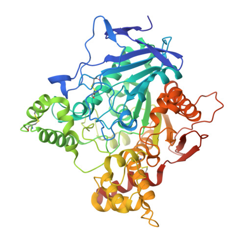 | |
UniProt | |||||
Entity Groups | |||||
| Sequence Clusters | 30% Identity50% Identity70% Identity90% Identity95% Identity100% Identity | ||||
| UniProt Group | P04058 | ||||
Glycosylation | |||||
| Glycosylation Sites: 1 | |||||
Sequence AnnotationsExpand | |||||
Reference Sequence | |||||
| Ligands 4 Unique | |||||
|---|---|---|---|---|---|
| ID | Chains | Name / Formula / InChI Key | 2D Diagram | 3D Interactions | |
| NAG Download:Ideal Coordinates CCD File | D [auth A], E [auth A] | 2-acetamido-2-deoxy-beta-D-glucopyranose C8 H15 N O6 OVRNDRQMDRJTHS-FMDGEEDCSA-N |  | ||
| PG4 Download:Ideal Coordinates CCD File | F [auth A] | TETRAETHYLENE GLYCOL C8 H18 O5 UWHCKJMYHZGTIT-UHFFFAOYSA-N |  | ||
| XE Download:Ideal Coordinates CCD File | B [auth A], C [auth A] | XENON Xe FHNFHKCVQCLJFQ-UHFFFAOYSA-N |  | ||
| PEG Download:Ideal Coordinates CCD File | G [auth A] H [auth A] I [auth A] J [auth A] K [auth A] | DI(HYDROXYETHYL)ETHER C4 H10 O3 MTHSVFCYNBDYFN-UHFFFAOYSA-N |  | ||
| Length ( Å ) | Angle ( ˚ ) |
|---|---|
| a = 111.769 | α = 90 |
| b = 111.769 | β = 90 |
| c = 137.232 | γ = 120 |
| Software Name | Purpose |
|---|---|
| MAR345dtb | data collection |
| HKL2Map | model building |
| SHELXL-97 | refinement |
| XDS | data reduction |
| XDS | data scaling |
| SHELX | phasing |


