


Crystal structure of Glutathione S Transferase in complex with glutathione from Pseudomonas fluorescens
Agarwal, R., Burley, S.K., Swaminathan, S.To be published.
Experimental Data Snapshot
Entity ID: 1 | |||||
|---|---|---|---|---|---|
| Molecule | Chains | Sequence Length | Organism | Details | Image |
| uncharacterized protein GST_N | 213 | Pseudomonas protegens Pf-5 | Mutation(s): 0 Gene Names: PFL_2580 | 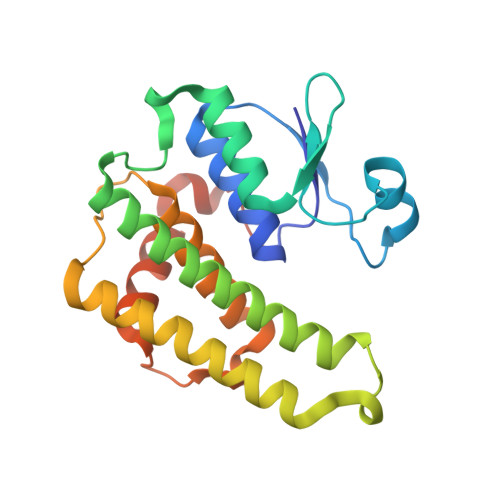 | |
UniProt | |||||
Entity Groups | |||||
| Sequence Clusters | 30% Identity50% Identity70% Identity90% Identity95% Identity100% Identity | ||||
| UniProt Group | Q4KDJ6 | ||||
Sequence AnnotationsExpand | |||||
Reference Sequence | |||||
| Ligands 1 Unique | |||||
|---|---|---|---|---|---|
| ID | Chains | Name / Formula / InChI Key | 2D Diagram | 3D Interactions | |
| GSH Download:Ideal Coordinates CCD File | C [auth A] | Glutathione C10 H17 N3 O6 S RWSXRVCMGQZWBV-WDSKDSINSA-N |  | ||
| Entity ID: 2 | |||||
|---|---|---|---|---|---|
| ID | Chains | Name | Type/Class | 2D Diagram | 3D Interactions |
| PRD_002593 (GSH) Query on PRD_002593 | C [auth A] | Glutathione | Peptide-like / Oxidation-reduction | | |
| Length ( Å ) | Angle ( ˚ ) |
|---|---|
| a = 48.521 | α = 90 |
| b = 57.866 | β = 106 |
| c = 74.796 | γ = 90 |
| Software Name | Purpose |
|---|---|
| HKL-2000 | data collection |
| SHELXD | phasing |
| SHARP | phasing |
| Coot | model building |
| CCP4 | refinement |
| CNS | refinement |
| HKL-2000 | data reduction |
| SCALEPACK | data scaling |


