3M0B | pdb_00003m0b

Ru-Porphyrin Protein Scaffolds for Sensing O2


Experimental Data Snapshot

  • Method: X-RAY DIFFRACTION
  • Resolution: 2.00 Å
  • R-Value Free: 
    0.225 (Depositor), 0.225 (DCC) 
  • R-Value Work: 
    0.204 (Depositor), 0.200 (DCC) 
  • R-Value Observed: 
    0.206 (Depositor) 

Starting Model: experimental
View more details

wwPDB Validation 3D Report Full Report

Validation slider image for 3M0B

Ligand Structure Quality Assessment 


This is version 1.2 of the entry. See complete history

Literature

Ru-porphyrin protein scaffolds for sensing O2.

Winter, M.B.McLaurin, E.J.Reece, S.Y.Olea, C.Nocera, D.G.Marletta, M.A.

(2010) J Am Chem Soc 132: 5582-5583

  • DOI: https://doi.org/10.1021/ja101527r
  • Primary Citation Related Structures: 
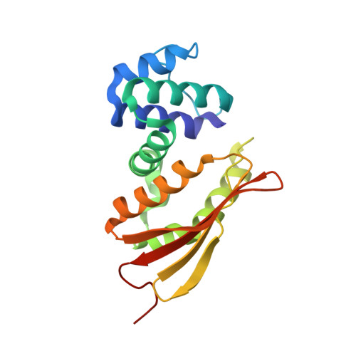
    3M0B

  • PubMed Abstract: 

    Hemoprotein-based scaffolds containing phosphorescent ruthenium(II) CO mesoporphyrin IX (RuMP) are reported here for oxygen (O(2)) sensing in biological contexts. RuMP was incorporated into the protein scaffolds during protein expression utilizing a novel method that we have described previously. A high-resolution (2.00 A) crystal structure revealed that the unnatural porphyrin binds to the proteins in a manner similar to the native heme and does not perturb the protein fold. The protein scaffolds were found to provide unique coordination environments for RuMP and modulate the porphyrin emission properties. Emission lifetime measurements demonstrate a linear O(2) response within the physiological range and precision comparable to commercial O(2) sensors. The RuMP proteins are robust, readily modifiable platforms and display promising O(2) sensing properties for future in vivo applications.


  • Organizational Affiliation
    • Department of Chemistry, QB3 Institute, and Division of Physical Biosciences, Lawrence Berkeley National Laboratory, University of California, Berkeley, California 94720-3220, USA.

Macromolecule Content 

  • Total Structure Weight: 22.74 kDa 
  • Atom Count: 1,646 
  • Modeled Residue Count: 185 
  • Deposited Residue Count: 188 
  • Unique protein chains: 1

Macromolecules

Find similar proteins by:|  3D Structure
Entity ID: 1
MoleculeChains  Sequence LengthOrganismDetailsImage
Methyl-accepting chemotaxis protein188Caldanaerobacter subterraneus subsp. tengcongensisMutation(s): 0 
UniProt
Find proteins for Q8RBX6 (Caldanaerobacter subterraneus subsp. tengcongensis (strain DSM 15242 / JCM 11007 / NBRC 100824 / MB4))
Explore Q8RBX6 
Go to UniProtKB:  Q8RBX6
Entity Groups
Sequence Clusters30% Identity50% Identity70% Identity90% Identity95% Identity100% Identity
UniProt GroupQ8RBX6
Sequence Annotations
Expand
Reference Sequence

Small Molecules

Ligands 2 Unique
IDChains Name / Formula / InChI Key2D Diagram3D Interactions
RUR

Query on RUR


Download:Ideal Coordinates CCD File
B [auth A][3,3'-(7,12-diethyl-3,8,13,17-tetramethylporphyrin-2,18-diyl-kappa~4~N~21~,N~22~,N~23~,N~24~)dipropanoato(2-)]ruthenium
C34 H36 N4 O4 Ru
UBGGULWHFXPIQZ-RGGAHWMASA-L
CMO

Query on CMO


Download:Ideal Coordinates CCD File
C [auth A]CARBON MONOXIDE
C O
UGFAIRIUMAVXCW-UHFFFAOYSA-N

Experimental Data & Validation

Experimental Data

  • Method: X-RAY DIFFRACTION
  • Resolution: 2.00 Å
  • R-Value Free:  0.225 (Depositor), 0.225 (DCC) 
  • R-Value Work:  0.204 (Depositor), 0.200 (DCC) 
  • R-Value Observed: 0.206 (Depositor) 
Space Group: P 61 2 2
Unit Cell:
Length ( Å )Angle ( ˚ )
a = 61.183α = 90
b = 61.183β = 90
c = 245.116γ = 120
Software Package:
Software NamePurpose
ADSCdata collection
PHASERphasing
PHENIXrefinement
HKL-2000data reduction
HKL-2000data scaling

Structure Validation

View Full Validation Report


Ligand Structure Quality Assessment 


Entry History 

Deposition Data

Revision History  (Full details and data files)

  • Version 1.0: 2010-04-14
    Type: Initial release
  • Version 1.1: 2011-07-13
    Changes: Version format compliance
  • Version 1.2: 2023-09-06
    Changes: Data collection, Database references, Derived calculations, Refinement description