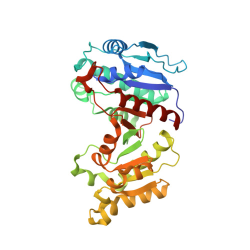
Crystal structure of 4-hydroxythreonine-4-phosphate dehydrogenase from Yersinia pestis CO92
Nocek, B., Maltseva, N., Kwon, K., Makowska-Grzyska, M., Anderson, W., Joachimiak, A., Center for Structural Genomics of Infectious Diseases (CSGID)To be published.