3LW2 | pdb_00003lw2

Mouse Plasminogen Activator Inhibitor-1 (PAI-1)


Experimental Data Snapshot

  • Method: X-RAY DIFFRACTION
  • Resolution: 1.93 Å
  • R-Value Free: 
    0.218 (Depositor), 0.225 (DCC) 
  • R-Value Work: 
    0.181 (Depositor) 
  • R-Value Observed: 
    0.183 (Depositor) 

Starting Model: experimental
View more details

wwPDB Validation 3D Report Full Report

Validation slider image for 3LW2

This is version 1.4 of the entry. See complete history

Literature

Subtle structural differences between human and mouse PAI-1 reveal the basis for biochemical differences.

Dewilde, M.Van De Craen, B.Compernolle, G.Madsen, J.B.Strelkov, S.Gils, A.Declerck, P.J.

(2010) J Struct Biol 171: 95-101

  • DOI: https://doi.org/10.1016/j.jsb.2010.03.006
  • Primary Citation Related Structures: 
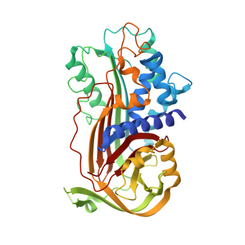
    3LW2

  • PubMed Abstract: 

    Plasminogen activator inhibitor-1 (PAI-1) is a serine protease inhibitor (serpin) that plays an important role in cardiovascular disorders and tumor development. The potential role of PAI-1 as a drug target has been evaluated in various animal models (e.g. mouse and rat). Sensitivity to PAI-1 inhibitory agents varied in different species. To date, absence of PAI-1 structures from species other than human hampers efforts to reveal the molecular basis for the observed species differences. Here we describe the structure of latent mouse PAI-1. Comparison with available structures of human PAI-1 reveals (1) a differential positioning of α-helix A; (2) differences in the gate region; and (3) differences in the reactive center loop position. We demonstrate that the optimal binding site of inhibitors may be dependent on the orthologs, and our results affect strategies in the rational design of a pharmacologically active PAI-1 inhibitor.


  • Organizational Affiliation
    • Laboratory for Pharmaceutical Biology, Katholieke Universiteit Leuven, Belgium.

Macromolecule Content 

  • Total Structure Weight: 42.8 kDa 
  • Atom Count: 3,153 
  • Modeled Residue Count: 365 
  • Deposited Residue Count: 379 
  • Unique protein chains: 1

Macromolecules

Find similar proteins by:|  3D Structure
Entity ID: 1
MoleculeChains  Sequence LengthOrganismDetailsImage
Plasminogen activator inhibitor 1379Mus musculusMutation(s): 0 
Gene Names: Serpine1Mr1Pai1Planh1
UniProt & NIH Common Fund Data Resources
Find proteins for P22777 (Mus musculus)
Explore P22777 
Go to UniProtKB:  P22777
IMPC:  MGI:97608
Entity Groups
Sequence Clusters30% Identity50% Identity70% Identity90% Identity95% Identity100% Identity
UniProt GroupP22777
Sequence Annotations
Expand
Reference Sequence

Experimental Data & Validation

Experimental Data

  • Method: X-RAY DIFFRACTION
  • Resolution: 1.93 Å
  • R-Value Free:  0.218 (Depositor), 0.225 (DCC) 
  • R-Value Work:  0.181 (Depositor) 
  • R-Value Observed: 0.183 (Depositor) 
Space Group: C 1 2 1
Unit Cell:
Length ( Å )Angle ( ˚ )
a = 150.9α = 90
b = 62.3β = 105.5
c = 52.2γ = 90
Software Package:
Software NamePurpose
XSCALEdata scaling
PHASERphasing
REFMACrefinement
PDB_EXTRACTdata extraction
XDSdata reduction

Structure Validation

View Full Validation Report


Entry History 

Deposition Data

Revision History  (Full details and data files)

  • Version 1.0: 2010-04-07
    Type: Initial release
  • Version 1.1: 2011-07-13
    Changes: Version format compliance
  • Version 1.2: 2013-06-19
    Changes: Database references
  • Version 1.3: 2017-11-08
    Changes: Refinement description
  • Version 1.4: 2023-09-06
    Changes: Data collection, Database references, Refinement description