3LVG

Crystal structure of a clathrin heavy chain and clathrin light chain complex


Experimental Data Snapshot

  • Method: X-RAY DIFFRACTION
  • Resolution: 7.94 Å
  • R-Value Free: 0.425 
  • R-Value Work: 0.419 

wwPDB Validation   3D Report Full Report


This is version 1.4 of the entry. See complete history


Literature

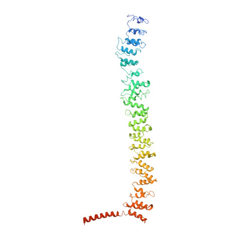
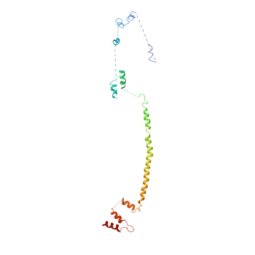
Conformation switching of clathrin light chain regulates clathrin lattice assembly.

Wilbur, J.D.Hwang, P.K.Ybe, J.A.Lane, M.Sellers, B.D.Jacobson, M.P.Fletterick, R.J.Brodsky, F.M.

(2010) Dev Cell 18: 841-848

  • DOI: https://doi.org/10.1016/j.devcel.2010.04.007
  • Primary Citation of Related Structures:  
    3LVG, 3LVH

  • PubMed Abstract: 

    Clathrin-coated vesicle formation is responsible for membrane traffic to and from the endocytic pathway during receptor-mediated endocytosis and organelle biogenesis, influencing how cells relate to their environment. Generating these vesicles involves self-assembly of clathrin molecules into a latticed coat on membranes that recruits receptors and organizes protein machinery necessary for budding. Here we define a molecular mechanism regulating clathrin lattice formation by obtaining structural information from co-crystals of clathrin subunits. Low resolution X-ray diffraction data (7.9-9.0 A) was analyzed using a combination of molecular replacement with an energy-minimized model and noncrystallographic symmetry averaging. Resulting topological information revealed two conformations of the regulatory clathrin light chain bound to clathrin heavy chain. Based on protein domain positions, mutagenesis, and biochemical assays, we identify an electrostatic interaction between the clathrin subunits that allows the observed conformational variation in clathrin light chains to alter the conformation of the clathrin heavy chain and thereby regulates assembly.


  • Organizational Affiliation

    Graduate Program in Biophysics, University of California, San Francisco, San Francisco, CA 94143, USA.


Macromolecules
Find similar proteins by:  (by identity cutoff)  |  3D Structure
Entity ID: 1
MoleculeChains Sequence LengthOrganismDetailsImage
Clathrin heavy chain 1
A, B, C
624Bos taurusMutation(s): 0 
Gene Names: CLTC
UniProt
Find proteins for P49951 (Bos taurus)
Explore P49951 
Go to UniProtKB:  P49951
Entity Groups  
Sequence Clusters30% Identity50% Identity70% Identity90% Identity95% Identity100% Identity
UniProt GroupP49951
Sequence Annotations
Expand
  • Reference Sequence
Find similar proteins by:  (by identity cutoff)  |  3D Structure
Entity ID: 2
MoleculeChains Sequence LengthOrganismDetailsImage
Clathrin light chain B
D, E, F
190Bos taurusMutation(s): 0 
Gene Names: CLTBCLTLB
UniProt
Find proteins for P04975 (Bos taurus)
Explore P04975 
Go to UniProtKB:  P04975
Entity Groups  
Sequence Clusters30% Identity50% Identity70% Identity90% Identity95% Identity100% Identity
UniProt GroupP04975
Sequence Annotations
Expand
  • Reference Sequence
Experimental Data & Validation

Experimental Data

  • Method: X-RAY DIFFRACTION
  • Resolution: 7.94 Å
  • R-Value Free: 0.425 
  • R-Value Work: 0.419 
  • Space Group: I 41 2 2
Unit Cell:
Length ( Å )Angle ( ˚ )
a = 228.56α = 90
b = 228.56β = 90
c = 710.32γ = 90
Software Package:
Software NamePurpose
DENZOdata reduction
SCALEPACKdata scaling
PHASERphasing
CNSrefinement
PDB_EXTRACTdata extraction

Structure Validation

View Full Validation Report


Entry History 

Deposition Data

Revision History  (Full details and data files)

  • Version 1.0: 2010-06-09
    Type: Initial release
  • Version 1.1: 2011-07-13
    Changes: Version format compliance
  • Version 1.2: 2017-08-02
    Changes: Refinement description, Source and taxonomy
  • Version 1.3: 2017-11-08
    Changes: Refinement description
  • Version 1.4: 2024-02-21
    Changes: Data collection, Database references