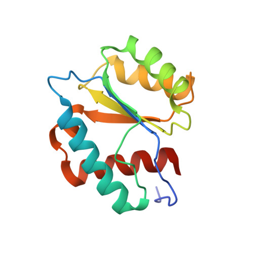
3LUG

Crystal structure of MID domain from hAGO2 in complex with CMP


Experimental Data Snapshot

  • Method: X-RAY DIFFRACTION
  • Resolution: 1.85 Å
  • R-Value Free: 0.225 
  • R-Value Work: 0.182 
  • R-Value Observed: 0.185 

wwPDB Validation   3D Report Full Report


Ligand Structure Quality Assessment 


This is version 1.3 of the entry. See complete history


Literature

Structural basis for 5'-nucleotide base-specific recognition of guide RNA by human AGO2.

Frank, F.Sonenberg, N.Nagar, B.

(2010) Nature 465: 818-822

  • DOI: https://doi.org/10.1038/nature09039
  • Primary Citation of Related Structures:  
    3LUC, 3LUD, 3LUG, 3LUH, 3LUJ, 3LUK

  • PubMed Abstract: 

    MicroRNAs (miRNAs) mediate post-transcriptional gene regulation through association with Argonaute proteins (AGOs). Crystal structures of archaeal and bacterial homologues of AGOs have shown that the MID (middle) domain mediates the interaction with the phosphorylated 5' end of the miRNA guide strand and this interaction is thought to be independent of the identity of the 5' nucleotide in these systems. However, analysis of the known sequences of eukaryotic miRNAs and co-immunoprecipitation experiments indicate that there is a clear bias for U or A at the 5' position. Here we report the crystal structure of a MID domain from a eukaryotic AGO protein, human AGO2. The structure, in complex with nucleoside monophosphates (AMP, CMP, GMP, and UMP) mimicking the 5' end of miRNAs, shows that there are specific contacts made between the base of UMP or AMP and a rigid loop in the MID domain. Notably, the structure of the loop discriminates against CMP and GMP and dissociation constants calculated from NMR titration experiments confirm these results, showing that AMP (0.26 mM) and UMP (0.12 mM) bind with up to 30-fold higher affinity than either CMP (3.6 mM) or GMP (3.3 mM). This study provides structural evidence for nucleotide-specific interactions in the MID domain of eukaryotic AGO proteins and explains the observed preference for U or A at the 5' end of miRNAs.


  • Organizational Affiliation

    Department of Biochemistry, McGill University, Montréal, Québec H3G 0B1, Canada.


Macromolecules
Find similar proteins by:  (by identity cutoff)  |  3D Structure
Entity ID: 1
MoleculeChains Sequence LengthOrganismDetailsImage
Protein argonaute-2
A, B, C
138Homo sapiensMutation(s): 0 
Gene Names: EIF2C2AGO2
UniProt & NIH Common Fund Data Resources
Find proteins for Q9UKV8 (Homo sapiens)
Explore Q9UKV8 
Go to UniProtKB:  Q9UKV8
PHAROS:  Q9UKV8
GTEx:  ENSG00000123908 
Entity Groups  
Sequence Clusters30% Identity50% Identity70% Identity90% Identity95% Identity100% Identity
UniProt GroupQ9UKV8
Sequence Annotations
Expand
  • Reference Sequence
Experimental Data & Validation

Experimental Data

  • Method: X-RAY DIFFRACTION
  • Resolution: 1.85 Å
  • R-Value Free: 0.225 
  • R-Value Work: 0.182 
  • R-Value Observed: 0.185 
  • Space Group: P 1
Unit Cell:
Length ( Å )Angle ( ˚ )
a = 40.503α = 86.06
b = 47.306β = 72.8
c = 66.403γ = 83.18
Software Package:
Software NamePurpose
PHENIXrefinement
CNSrefinement
HKL-2000data collection
HKL-2000data reduction
HKL-2000data scaling
CNSphasing

Structure Validation

View Full Validation Report


Ligand Structure Quality Assessment 


Entry History 

Deposition Data

Revision History  (Full details and data files)

  • Version 1.0: 2010-05-26
    Type: Initial release
  • Version 1.1: 2011-07-13
    Changes: Version format compliance
  • Version 1.2: 2019-07-17
    Changes: Data collection, Refinement description
  • Version 1.3: 2024-02-21
    Changes: Data collection, Database references, Derived calculations