3LT3

Crystal structure of Rv3671c from M. tuberculosis H37Rv, Ser343Ala mutant, inactive form


Experimental Data Snapshot

  • Method: X-RAY DIFFRACTION
  • Resolution: 2.10 Å
  • R-Value Free: 0.276 
  • R-Value Work: 0.227 
  • R-Value Observed: 0.229 

wwPDB Validation   3D Report Full Report


This is version 1.2 of the entry. See complete history


Literature

Structural insight into serine protease Rv3671c that Protects M. tuberculosis from oxidative and acidic stress.

Biswas, T.Small, J.Vandal, O.Odaira, T.Deng, H.Ehrt, S.Tsodikov, O.V.

(2010) Structure 18: 1353-1363

  • DOI: https://doi.org/10.1016/j.str.2010.06.017
  • Primary Citation of Related Structures:  
    3K6Y, 3K6Z, 3LT3

  • PubMed Abstract: 

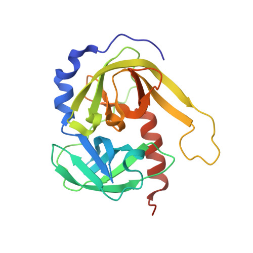
    Rv3671c, a putative serine protease, is crucial for persistence of Mycobacterium tuberculosis in the hostile environment of the phagosome. We show that Rv3671c is required for M. tuberculosis resistance to oxidative stress in addition to its role in protection from acidification. Structural and biochemical analyses demonstrate that the periplasmic domain of Rv3671c is a functional serine protease of the chymotrypsin family and, remarkably, that its activity increases on oxidation. High-resolution crystal structures of this protease in an active strained state and in an inactive relaxed state reveal that a solvent-exposed disulfide bond controls the protease activity by constraining two distant regions of Rv3671c and stabilizing it in the catalytically active conformation. In vitro biochemical studies confirm that activation of the protease in an oxidative environment is dependent on this reversible disulfide bond. These results suggest that the disulfide bond modulates activity of Rv3671c depending on the oxidative environment in vivo.


  • Organizational Affiliation

    Department of Medicinal Chemistry, College of Pharmacy, University of Michigan, Ann Arbor, MI 48109, USA.


Macromolecules
Find similar proteins by:  (by identity cutoff)  |  3D Structure
Entity ID: 1
MoleculeChains Sequence LengthOrganismDetailsImage
POSSIBLE MEMBRANE-ASSOCIATED SERINE PROTEASE
A, B
217Mycobacterium tuberculosis H37RvMutation(s): 1 
Gene Names: MT3772Rv3671c
EC: 3.4.21
UniProt
Find proteins for P9WHR9 (Mycobacterium tuberculosis (strain ATCC 25618 / H37Rv))
Explore P9WHR9 
Go to UniProtKB:  P9WHR9
Entity Groups  
Sequence Clusters30% Identity50% Identity70% Identity90% Identity95% Identity100% Identity
UniProt GroupP9WHR9
Sequence Annotations
Expand
  • Reference Sequence
Experimental Data & Validation

Experimental Data

  • Method: X-RAY DIFFRACTION
  • Resolution: 2.10 Å
  • R-Value Free: 0.276 
  • R-Value Work: 0.227 
  • R-Value Observed: 0.229 
  • Space Group: P 1
Unit Cell:
Length ( Å )Angle ( ˚ )
a = 42.603α = 101.68
b = 44.207β = 97.56
c = 53.356γ = 105.05
Software Package:
Software NamePurpose
REFMACrefinement
HKL-2000data reduction
HKL-2000data scaling
PHASERphasing

Structure Validation

View Full Validation Report


Entry History 

Deposition Data

Revision History  (Full details and data files)

  • Version 1.0: 2010-11-03
    Type: Initial release
  • Version 1.1: 2011-07-13
    Changes: Version format compliance
  • Version 1.2: 2023-09-06
    Changes: Data collection, Database references, Refinement description