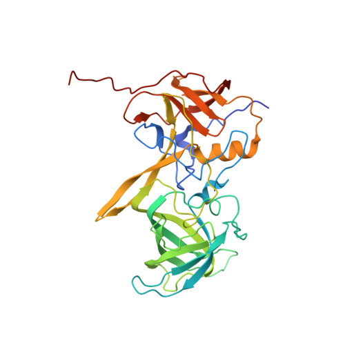
High-resolution x-ray structure and functional analysis of the murine norovirus 1 capsid protein protruding domain.
Taube, S., Rubin, J.R., Katpally, U., Smith, T.J., Kendall, A., Stuckey, J.A., Wobus, C.E.(2010) J Virol 84: 5695-5705
- PubMed: 20335262 Search on PubMedSearch on PubMed Central
- DOI: https://doi.org/10.1128/JVI.00316-10
- Primary Citation Related Structures:
3LQ6, 3LQE - PubMed Abstract:
Murine noroviruses (MNV) are closely related to the human noroviruses (HuNoV), which cause the majority of nonbacterial gastroenteritis. Unlike HuNoV, MNV grow in culture and in a small-animal model that represents a tractable model to study norovirus biology. To begin a detailed investigation of molecular events that occur during norovirus binding to cells, the crystallographic structure of the murine norovirus 1 (MNV-1) capsid protein protruding (P) domain has been determined. Crystallization of the bacterially expressed protein yielded two different crystal forms (Protein Data Bank identifiers [PDB ID], 3LQ6 and 3LQE). Comparison of the structures indicated a large degree of structural mobility in loops on the surface of the P2 subdomain. Specifically, the A'-B' and E'-F' loops were found in open and closed conformations. These regions of high mobility include the known escape mutation site for the neutralizing antibody A6.2 and an attenuation mutation site, which arose after serial passaging in culture and led to a loss in lethality in STAT1(-/-) mice, respectively. Modeling of a Fab fragment and crystal structures of the P dimer into the cryoelectron microscopy three-dimensional (3D) image reconstruction of the A6.2/MNV-1 complex indicated that the closed conformation is most likely bound to the Fab fragment and that the antibody contact is localized to the A'-B' and E'-F' loops. Therefore, we hypothesize that these loop regions and the flexibility of the P domains play important roles during MNV-1 binding to the cell surface.
- Department of Microbiology and Immunology, University of Michigan Medical School, Ann Arbor, Michigan 48109, USA.
Organizational Affiliation: