The Subtilisin-Like Protease AprV2 Is Required for Virulence and Uses a Novel Disulphide-Tethered Exosite to Bind Substrates
Kennan, R.M., Wong, W., Dhungyel, O.P., Han, X., Wong, D., Parker, D., Rosado, C.J., Law, R.H.P., McGowan, S., Reeve, S.B., Levina, V., Powers, G.A., Pike, R.N., Bottomley, S.P., Smith, A.I., Marsh, I., Whittington, R.J., Whisstock, J.C., Porter, C.J., Rood, J.I.(2010) PLoS Pathog 6: e1001210-e1001210
- PubMed: 21124876 Search on PubMedSearch on PubMed Central
- DOI: https://doi.org/10.1371/journal.ppat.1001210
- Primary Citation Related Structures:
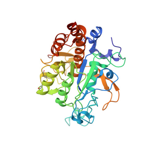
3LPA, 3LPC, 3LPD - PubMed Abstract:
Many bacterial pathogens produce extracellular proteases that degrade the extracellular matrix of the host and therefore are involved in disease pathogenesis. Dichelobacter nodosus is the causative agent of ovine footrot, a highly contagious disease that is characterized by the separation of the hoof from the underlying tissue. D. nodosus secretes three subtilisin-like proteases whose analysis forms the basis of diagnostic tests that differentiate between virulent and benign strains and have been postulated to play a role in virulence. We have constructed protease mutants of D. nodosus; their analysis in a sheep virulence model revealed that one of these enzymes, AprV2, was required for virulence. These studies challenge the previous hypothesis that the elastase activity of AprV2 is important for disease progression, since aprV2 mutants were virulent when complemented with aprB2, which encodes a variant that has impaired elastase activity. We have determined the crystal structures of both AprV2 and AprB2 and characterized the biological activity of these enzymes. These data reveal that an unusual extended disulphide-tethered loop functions as an exosite, mediating effective enzyme-substrate interactions. The disulphide bond and Tyr92, which was located at the exposed end of the loop, were functionally important. Bioinformatic analyses suggested that other pathogenic bacteria may have proteases that utilize a similar mechanism. In conclusion, we have used an integrated multidisciplinary combination of bacterial genetics, whole animal virulence trials in the original host, biochemical studies, and comprehensive analysis of crystal structures to provide the first definitive evidence that the extracellular secreted proteases produced by D. nodosus are required for virulence and to elucidate the molecular mechanism by which these proteases bind to their natural substrates. We postulate that this exosite mechanism may be used by proteases produced by other bacterial pathogens of both humans and animals.
- Monash University, Clayton, Victoria, Australia.
Organizational Affiliation: