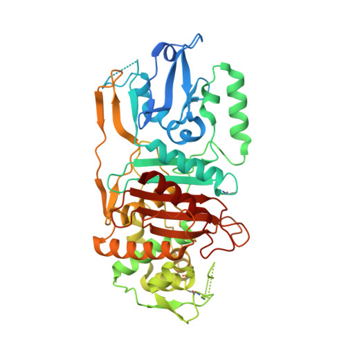
Unusual conformation of the SxN motif in the crystal structure of penicillin-binding protein A from Mycobacterium tuberculosis.
Fedarovich, A., Nicholas, R.A., Davies, C.(2010) J Mol Biology 398: 54-65
- PubMed: 20206184 Search on PubMedSearch on PubMed Central
- DOI: https://doi.org/10.1016/j.jmb.2010.02.046
- Primary Citation Related Structures:
3LO7 - PubMed Abstract:
PBPA from Mycobacterium tuberculosis is a class B-like penicillin-binding protein (PBP) that is not essential for cell growth in M. tuberculosis, but is important for proper cell division in Mycobacterium smegmatis. We have determined the crystal structure of PBPA at 2.05 A resolution, the first published structure of a PBP from this important pathogen. Compared to other PBPs, PBPA has a relatively small N-terminal domain, and conservation of a cluster of charged residues within this domain suggests that PBPA is more related to class B PBPs than previously inferred from sequence analysis. The C-terminal domain is a typical transpeptidase fold and contains the three conserved active-site motifs characteristic of penicillin-interacting enzymes. Whilst the arrangement of the SxxK and KTG motifs is similar to that observed in other PBPs, the SxN motif is markedly displaced away from the active site, such that its serine (Ser281) is not involved in hydrogen bonding with residues of the other two motifs. A disulfide bridge between Cys282 (the "x" of the SxN motif) and Cys266, which resides on an adjacent loop, may be responsible for this unusual conformation. Another interesting feature of the structure is a relatively long connection between beta 5 and alpha 11, which restricts the space available in the active site of PBPA and suggests that conformational changes would be required to accommodate peptide substrate or beta-lactam antibiotics during acylation. Finally, the structure shows that one of the two threonines postulated to be targets for phosphorylation is inaccessible (Thr362), whereas the other (Thr437) is well placed on a surface loop near the active site.
- Department of Biochemistry and Molecular Biology, Medical University of South Carolina, 173 Ashley Avenue, Charleston, SC 29425, USA.
Organizational Affiliation: