


Crystal structure of ParE subunit
Heo, Y.-S.To be published.
Experimental Data Snapshot
wwPDB Validation 3D Report Full Report

Entity ID: 1 | |||||
|---|---|---|---|---|---|
| Molecule | Chains | Sequence Length | Organism | Details | Image |
| Topoisomerase IV subunit B | 408 | Xanthomonas oryzae pv. oryzae KACC 10331 | Mutation(s): 0 Gene Names: parE, XOO2969 EC: 5.99.1.3 (PDB Primary Data), 5.6.2.2 (UniProt) | 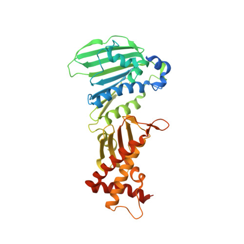 | |
UniProt | |||||
Entity Groups | |||||
| Sequence Clusters | 30% Identity50% Identity70% Identity90% Identity95% Identity100% Identity | ||||
| UniProt Group | Q5GYJ8 | ||||
Sequence AnnotationsExpand | |||||
Reference Sequence | |||||
| Length ( Å ) | Angle ( ˚ ) |
|---|---|
| a = 105.302 | α = 90 |
| b = 105.302 | β = 90 |
| c = 133.765 | γ = 90 |
| Software Name | Purpose |
|---|---|
| HKL-2000 | data collection |
| CNS | refinement |
| HKL-2000 | data reduction |
| HKL-2000 | data scaling |
| CNS | phasing |


