3LNF

Crystal structure of E-cadherin EC12 K14EW2A


Experimental Data Snapshot

  • Method: X-RAY DIFFRACTION
  • Resolution: 2.50 Å
  • R-Value Free: 0.241 
  • R-Value Work: 0.184 
  • R-Value Observed: 0.187 

wwPDB Validation   3D Report Full Report


This is version 1.3 of the entry. See complete history


Literature

Two-step adhesive binding by classical cadherins.

Harrison, O.J.Bahna, F.Katsamba, P.S.Jin, X.Brasch, J.Vendome, J.Ahlsen, G.Carroll, K.J.Price, S.R.Honig, B.Shapiro, L.

(2010) Nat Struct Mol Biol 17: 348-357

  • DOI: https://doi.org/10.1038/nsmb.1784
  • Primary Citation of Related Structures:  
    3LND, 3LNE, 3LNF, 3LNG, 3LNH, 3LNI

  • PubMed Abstract: 

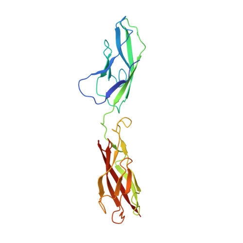
    Crystal structures of classical cadherins have revealed two dimeric configurations. In the first, N-terminal beta-strands of EC1 domains 'swap' between partner molecules. The second configuration (the 'X dimer'), also observed for T-cadherin, is mediated by residues near the EC1-EC2 calcium binding sites, and N-terminal beta-strands of partner EC1 domains, though held adjacent, do not swap. Here we show that strand-swapping mutants of type I and II classical cadherins form X dimers. Mutant cadherins impaired for X-dimer formation show no binding in short-time frame surface plasmon resonance assays, but in long-time frame experiments, they have homophilic binding affinities close to that of wild type. Further experiments show that exchange between monomers and dimers is slowed in these mutants. These results reconcile apparently disparate results from prior structural studies and suggest that X dimers are binding intermediates that facilitate the formation of strand-swapped dimers.


  • Organizational Affiliation

    Department of Biochemistry and Molecular Biophysics, Columbia University, New York, New York, USA.


Macromolecules
Find similar proteins by:  (by identity cutoff)  |  3D Structure
Entity ID: 1
MoleculeChains Sequence LengthOrganismDetailsImage
Cadherin-1
A, B
213Mus musculusMutation(s): 2 
Gene Names: Cdh1
UniProt & NIH Common Fund Data Resources
Find proteins for P09803 (Mus musculus)
Explore P09803 
Go to UniProtKB:  P09803
IMPC:  MGI:88354
Entity Groups  
Sequence Clusters30% Identity50% Identity70% Identity90% Identity95% Identity100% Identity
UniProt GroupP09803
Sequence Annotations
Expand
  • Reference Sequence
Experimental Data & Validation

Experimental Data

  • Method: X-RAY DIFFRACTION
  • Resolution: 2.50 Å
  • R-Value Free: 0.241 
  • R-Value Work: 0.184 
  • R-Value Observed: 0.187 
  • Space Group: P 1
Unit Cell:
Length ( Å )Angle ( ˚ )
a = 45.107α = 70.58
b = 47.383β = 88.13
c = 69.897γ = 75.5
Software Package:
Software NamePurpose
ADSCdata collection
MOLREPphasing
PHENIXrefinement
DENZOdata reduction
SCALEPACKdata scaling

Structure Validation

View Full Validation Report


Entry History 

Deposition Data

Revision History  (Full details and data files)

  • Version 1.0: 2010-03-02
    Type: Initial release
  • Version 1.1: 2011-07-13
    Changes: Version format compliance
  • Version 1.2: 2021-10-13
    Changes: Database references, Derived calculations
  • Version 1.3: 2023-09-06
    Changes: Data collection, Refinement description