3LNB

Crystal Structure Analysis of Arylamine N-acetyltransferase C from Bacillus anthracis


Experimental Data Snapshot

  • Method: X-RAY DIFFRACTION
  • Resolution: 2.01 Å
  • R-Value Free: 0.244 
  • R-Value Work: 0.203 

wwPDB Validation   3D Report Full Report


Ligand Structure Quality Assessment 


This is version 1.3 of the entry. See complete history


Literature

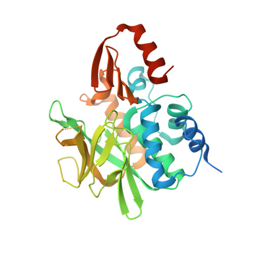
The Bacillus anthracis arylamine N-acetyltransferase ((BACAN)NAT1) that inactivates sulfamethoxazole, reveals unusual structural features compared with the other NAT isoenzymes.

Pluvinage, B.Li de la Sierra-Gallay, I.Kubiak, X.Xu, X.Dairou, J.Dupret, J.M.Rodrigues-Lima, F.

(2011) FEBS Lett 585: 3947-3952

  • DOI: https://doi.org/10.1016/j.febslet.2011.10.041
  • Primary Citation of Related Structures:  
    3LNB

  • PubMed Abstract: 

    Arylamine N-acetyltransferases (NATs) are xenobiotic-metabolizing enzymes that biotransform arylamine drugs. The Bacillus anthracis (BACAN)NAT1 enzyme affords increased resistance to the antibiotic sulfamethoxazole through its acetylation. We report the structure of (BACAN)NAT1. Unexpectedly, endogenous coenzymeA was present in the active site. The structure suggests that, contrary to the other prokaryotic NATs, (BACAN)NAT1 possesses a 14-residue insertion equivalent to the "mammalian insertion", a structural feature considered unique to mammalian NATs. Moreover, (BACAN)NAT1 structure shows marked differences in the mode of binding and location of coenzymeA when compared to the other NATs. This suggests that the mechanisms of cofactor recognition by NATs is more diverse than expected and supports the cofactor-binding site as being a unique subsite to target in drug design against bacterial NATs.


  • Organizational Affiliation

    Université Paris Diderot, Sorbonne Paris Cité, Unité BFA, EAC-CNRS 4413, Paris, France.


Macromolecules
Find similar proteins by:  (by identity cutoff)  |  3D Structure
Entity ID: 1
MoleculeChains Sequence LengthOrganismDetailsImage
N-acetyltransferase family protein309Bacillus anthracisMutation(s): 1 
EC: 2.3.1.5
UniProt
Find proteins for A0A6L7HKL6 (Bacillus anthracis)
Explore A0A6L7HKL6 
Go to UniProtKB:  A0A6L7HKL6
Entity Groups  
Sequence Clusters30% Identity50% Identity70% Identity90% Identity95% Identity100% Identity
UniProt GroupA0A6L7HKL6
Sequence Annotations
Expand
  • Reference Sequence
Experimental Data & Validation

Experimental Data

  • Method: X-RAY DIFFRACTION
  • Resolution: 2.01 Å
  • R-Value Free: 0.244 
  • R-Value Work: 0.203 
  • Space Group: P 41 21 2
Unit Cell:
Length ( Å )Angle ( ˚ )
a = 53.99α = 90
b = 53.99β = 90
c = 172.44γ = 90
Software Package:
Software NamePurpose
CNSrefinement
PDB_EXTRACTdata extraction
MxCuBEdata collection
XDSdata reduction
XSCALEdata scaling
CNSphasing

Structure Validation

View Full Validation Report


Ligand Structure Quality Assessment 


Entry History 

Deposition Data

Revision History  (Full details and data files)

  • Version 1.0: 2011-01-26
    Type: Initial release
  • Version 1.1: 2011-07-13
    Changes: Version format compliance
  • Version 1.2: 2021-02-03
    Changes: Database references, Derived calculations
  • Version 1.3: 2023-11-01
    Changes: Data collection, Database references, Refinement description