3LJ8 | pdb_00003lj8

Crystal Structure of MKP-4


Experimental Data Snapshot

  • Method: X-RAY DIFFRACTION
  • Resolution: 2.70 Å
  • R-Value Free: 
    0.304 (Depositor), 0.308 (DCC) 
  • R-Value Work: 
    0.238 (Depositor), 0.243 (DCC) 
  • R-Value Observed: 
    0.241 (Depositor) 

Starting Model: experimental
View more details

wwPDB Validation 3D Report Full Report

Validation slider image for 3LJ8

This is version 1.2 of the entry. See complete history

Literature

Exploring binding sites other than the catalytic core in the crystal structure of the catalytic domain of MKP-4

Jeong, D.G.Yoon, T.S.Jung, S.-K.Park, B.C.Park, H.S.Ryu, S.E.Kim, S.J.

(2011) Acta Crystallogr D Biol Crystallogr 67: 25-31

  • DOI: https://doi.org/10.1107/S0907444910042381
  • Primary Citation Related Structures: 
    3LJ8

  • PubMed Abstract: 

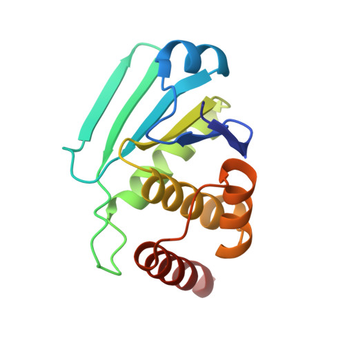
    Map kinase phosphatase 4 (MKP-4), which has been implicated in signalling pathways that negatively regulate glucose uptake, belongs to the dual-specificity phosphatase (DUSP) family. An inherent property of MKPs is an ability to undergo structural rearrangement, transitioning from a partially active to a fully active conformation. Here, a 2.7 Å resolution crystal structure of the catalytic domain of MKP-4 (MKP-4C) is presented. It was determined that the MKP-4C structure seriously deviates from canonical conformations of DUSPs and this characteristic feature results in significant gaps between the catalytic core and several surrounding loops which are unique compared with other MKP counterparts that adopt an active conformation. Using virtual library screening, it was found that inhibitors bind to MKP-4C with high affinity near these gaps. Inhibitors that target other binding sites instead of the active site can be utilized to prevent transition to a fully active conformation. Compounds that are able to make contacts with these sites in MKP-4 would not only provide a beneficial increase in affinity but may also permit greater specificity relative to other protein tyrosine phosphatases.


  • Organizational Affiliation
    • Medical Proteomics Research Center, Korea Research Institute of Bioscience and Biotechnology, Yuseong-Gu, Daejeon, Republic of Korea.

Macromolecule Content 

  • Total Structure Weight: 16.65 kDa 
  • Atom Count: 1,175 
  • Modeled Residue Count: 146 
  • Deposited Residue Count: 146 
  • Unique protein chains: 1

Macromolecules

Find similar proteins by:|  3D Structure
Entity ID: 1
MoleculeChains  Sequence LengthOrganismDetailsImage
Tyrosine-protein phosphatase146Homo sapiensMutation(s): 0 
EC: 3.1.3.48 (PDB Primary Data), 3.1.3.16 (PDB Primary Data)
UniProt & NIH Common Fund Data Resources
Find proteins for Q99956 (Homo sapiens)
Explore Q99956 
Go to UniProtKB:  Q99956
PHAROS:  Q99956
GTEx:  ENSG00000130829 
Entity Groups
Sequence Clusters30% Identity50% Identity70% Identity90% Identity95% Identity100% Identity
UniProt GroupQ99956
Sequence Annotations
Expand
Reference Sequence

Experimental Data & Validation

Experimental Data

  • Method: X-RAY DIFFRACTION
  • Resolution: 2.70 Å
  • R-Value Free:  0.304 (Depositor), 0.308 (DCC) 
  • R-Value Work:  0.238 (Depositor), 0.243 (DCC) 
  • R-Value Observed: 0.241 (Depositor) 
Space Group: P 32 2 1
Unit Cell:
Length ( Å )Angle ( ˚ )
a = 91.926α = 90
b = 91.926β = 90
c = 56.605γ = 120
Software Package:
Software NamePurpose
HKL-3000data collection
PHASERphasing
CNSrefinement
MOSFLMdata reduction
SCALAdata scaling

Structure Validation

View Full Validation Report


Entry History 

Deposition Data

Revision History  (Full details and data files)

  • Version 1.0: 2010-12-29
    Type: Initial release
  • Version 1.1: 2011-07-13
    Changes: Version format compliance
  • Version 1.2: 2023-11-01
    Changes: Data collection, Database references, Refinement description