3LIM | pdb_00003lim

Crystal structure of the pore forming toxin frac from sea anemone actinia fragacea


Experimental Data Snapshot

  • Method: X-RAY DIFFRACTION
  • Resolution: 1.80 Å
  • R-Value Free: 
    0.224 (Depositor), 0.227 (DCC) 
  • R-Value Work: 
    0.189 (Depositor) 
  • R-Value Observed: 
    0.191 (Depositor) 

wwPDB Validation 3D Report Full Report

Validation slider image for 3LIM

Ligand Structure Quality Assessment 


This is version 1.3 of the entry. See complete history

Literature

Structural insights into the oligomerization and architecture of eukaryotic membrane pore-forming toxins.

Mechaly, A.E.Bellomio, A.Gil-Carton, D.Morante, K.Valle, M.Gonzalez-Manas, J.M.Guerin, D.M.

(2011) Structure 19: 181-191

  • DOI: https://doi.org/10.1016/j.str.2010.11.013
  • Primary Citation Related Structures: 
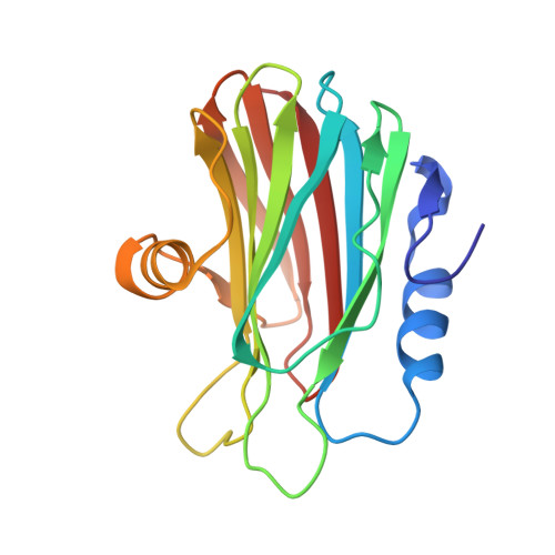
    3LIM

  • PubMed Abstract: 

    Pore-forming toxins (PFTs) are proteins that are secreted as soluble molecules and are inserted into membranes to form oligomeric transmembrane pores. In this paper, we report the crystal structure of Fragaceatoxin C (FraC), a PFT isolated from the sea anemone Actinia fragacea, at 1.8 Å resolution. It consists of a crown-shaped nonamer with an external diameter of about 11.0 nm and an internal diameter of approximately 5.0 nm. Cryoelectron microscopy studies of FraC in lipid bilayers reveal the pore structure that traverses the membrane. The shape and dimensions of the crystallographic oligomer are fully consistent with the membrane pore. The FraC structure provides insight into the interactions governing the assembly process and suggests the structural changes that allow for membrane insertion. We propose a nonameric pore model that spans the membrane by forming a lipid-free α-helical bundle pore.


  • Organizational Affiliation
    • Unidad de Biofísica (CSIC-UPV/EHU), Universidad del País Vasco, Barrio Sarriena S/N, 48940, Leioa, Vizcaya, Spain.

Macromolecule Content 

  • Total Structure Weight: 122.08 kDa 
  • Atom Count: 9,549 
  • Modeled Residue Count: 1,068 
  • Deposited Residue Count: 1,068 
  • Unique protein chains: 1

Macromolecules

Find similar proteins by:|  3D Structure
Entity ID: 1
MoleculeChains  Sequence LengthOrganismDetailsImage
Fragaceatoxin C
A, B, C, D, E
A, B, C, D, E, F
178Actinia fragaceaMutation(s): 0 
Membrane Entity: Yes 
UniProt
Find proteins for B9W5G6 (Actinia fragacea)
Explore B9W5G6 
Go to UniProtKB:  B9W5G6
Entity Groups
Sequence Clusters30% Identity50% Identity70% Identity90% Identity95% Identity100% Identity
UniProt GroupB9W5G6
Sequence Annotations
Expand
Reference Sequence

Small Molecules

Ligands 1 Unique
IDChains Name / Formula / InChI Key2D Diagram3D Interactions
LDA

Query on LDA


Download:Ideal Coordinates CCD File
G [auth A]
H [auth A]
I [auth A]
J [auth B]
K [auth B]
G [auth A],
H [auth A],
I [auth A],
J [auth B],
K [auth B],
L [auth B],
M [auth C],
N [auth C],
O [auth C],
P [auth D],
Q [auth D],
R [auth D],
S [auth E],
T [auth E],
U [auth E],
V [auth F],
W [auth F],
X [auth F]
LAURYL DIMETHYLAMINE-N-OXIDE
C14 H31 N O
SYELZBGXAIXKHU-UHFFFAOYSA-N

Experimental Data & Validation

Experimental Data

  • Method: X-RAY DIFFRACTION
  • Resolution: 1.80 Å
  • R-Value Free:  0.224 (Depositor), 0.227 (DCC) 
  • R-Value Work:  0.189 (Depositor) 
  • R-Value Observed: 0.191 (Depositor) 
Space Group: P 63 2 2
Unit Cell:
Length ( Å )Angle ( ˚ )
a = 117.899α = 90
b = 117.899β = 90
c = 343.228γ = 120
Software Package:
Software NamePurpose
PHASERphasing
PHENIXrefinement
MOSFLMdata reduction
SCALAdata scaling

Structure Validation

View Full Validation Report


Ligand Structure Quality Assessment 


Entry History 

Deposition Data

Revision History  (Full details and data files)

  • Version 1.0: 2010-12-15
    Type: Initial release
  • Version 1.1: 2011-07-13
    Changes: Version format compliance
  • Version 1.2: 2011-07-20
    Changes: Database references
  • Version 1.3: 2024-02-21
    Changes: Data collection, Database references, Derived calculations