3LH9

Crystal structure of mouse VPS26B(L197S/R199E) in spacegroup P41 21 2


Experimental Data Snapshot

  • Method: X-RAY DIFFRACTION
  • Resolution: 2.40 Å
  • R-Value Free: 0.228 
  • R-Value Work: 0.199 
  • R-Value Observed: 0.200 

wwPDB Validation   3D Report Full Report


This is version 1.3 of the entry. See complete history


Literature

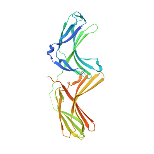
Assembly and solution structure of the core retromer protein complex.

Norwood, S.J.Shaw, D.J.Cowieson, N.P.Owen, D.J.Teasdale, R.D.Collins, B.M.

(2011) Traffic 12: 56-71

  • DOI: https://doi.org/10.1111/j.1600-0854.2010.01124.x
  • Primary Citation of Related Structures:  
    3LH8, 3LH9, 3LHA

  • PubMed Abstract: 

    Retromer is a peripheral membrane protein complex that has pleiotropic roles in endosomal membrane trafficking. The core of retromer possesses three subunits, VPS35, VPS29 and VPS26, that play different roles in binding to cargo, regulatory proteins and complex stabilization. We have performed an investigation of the thermodynamics of core retromer assembly using isothermal titration calorimetry (ITC) demonstrating that VPS35 acts as the central subunit to which VPS29 and VPS26 bind independently. Furthermore, we confirm that the conserved PRLYL motif of the large VPS35 subunit is critical for direct VPS26 interaction. Heat capacity measurements of VPS29 and VPS26 binding to VPS35 indicate extensive binding interfaces and suggest conformational alterations in VPS29 or VPS35 upon complex formation. Solution studies of the retromer core using small-angle X-ray scattering allow us to propose a model whereby VPS35 forms an extended platform with VPS29 and VPS26 bound at distal ends, with the potential for forming dimeric assemblies.


  • Organizational Affiliation

    Institute for Molecular Bioscience, The University of Queensland, St. Lucia, Queensland 4072, Australia.


Macromolecules
Find similar proteins by:  (by identity cutoff)  |  3D Structure
Entity ID: 1
MoleculeChains Sequence LengthOrganismDetailsImage
Vacuolar protein sorting-associated protein 26B
A, B
340Mus musculusMutation(s): 2 
Gene Names: Vps26b
UniProt
Find proteins for Q8C0E2 (Mus musculus)
Explore Q8C0E2 
Go to UniProtKB:  Q8C0E2
Entity Groups  
Sequence Clusters30% Identity50% Identity70% Identity90% Identity95% Identity100% Identity
UniProt GroupQ8C0E2
Sequence Annotations
Expand
  • Reference Sequence
Experimental Data & Validation

Experimental Data

  • Method: X-RAY DIFFRACTION
  • Resolution: 2.40 Å
  • R-Value Free: 0.228 
  • R-Value Work: 0.199 
  • R-Value Observed: 0.200 
  • Space Group: P 41 21 2
Unit Cell:
Length ( Å )Angle ( ˚ )
a = 118.35α = 90
b = 118.35β = 90
c = 214.513γ = 90
Software Package:
Software NamePurpose
PHASERphasing
PHENIXrefinement
MOSFLMdata reduction
SCALAdata scaling

Structure Validation

View Full Validation Report


Entry History 

Deposition Data

Revision History  (Full details and data files)

  • Version 1.0: 2010-02-02
    Type: Initial release
  • Version 1.1: 2011-07-13
    Changes: Version format compliance
  • Version 1.2: 2021-10-13
    Changes: Database references
  • Version 1.3: 2023-09-06
    Changes: Data collection, Refinement description