3LGF | pdb_00003lgf

Crystal structure of the 53BP1 tandem tudor domain in complex with p53K370me2


Experimental Data Snapshot

  • Method: X-RAY DIFFRACTION
  • Resolution: 1.50 Å
  • R-Value Free: 
    0.232 (Depositor), 0.230 (DCC) 
  • R-Value Work: 
    0.217 (Depositor), 0.212 (DCC) 
  • R-Value Observed: 
    0.217 (Depositor) 

Starting Model: experimental
View more details

wwPDB Validation 3D Report Full Report

Validation slider image for 3LGF

This is version 1.2 of the entry. See complete history

Literature

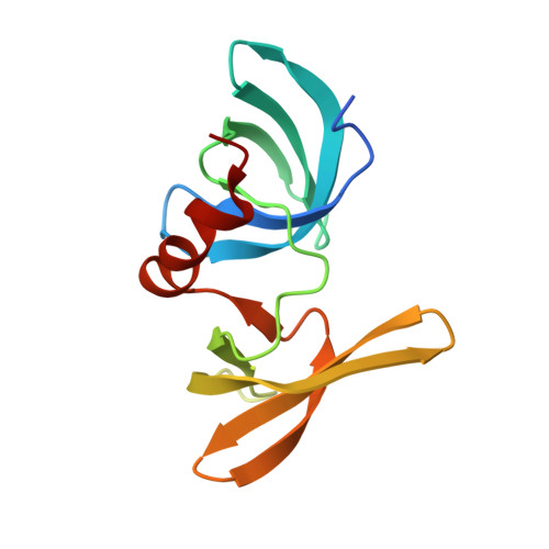
Structural insight into p53 recognition by the 53BP1 tandem Tudor domain.

Roy, S.Musselman, C.A.Kachirskaia, I.Hayashi, R.Glass, K.C.Nix, J.C.Gozani, O.Appella, E.Kutateladze, T.G.

(2010) J Mol Biology 398: 489-496

  • DOI: https://doi.org/10.1016/j.jmb.2010.03.024
  • Primary Citation Related Structures: 
    3LGF, 3LGL, 3LH0

  • PubMed Abstract: 

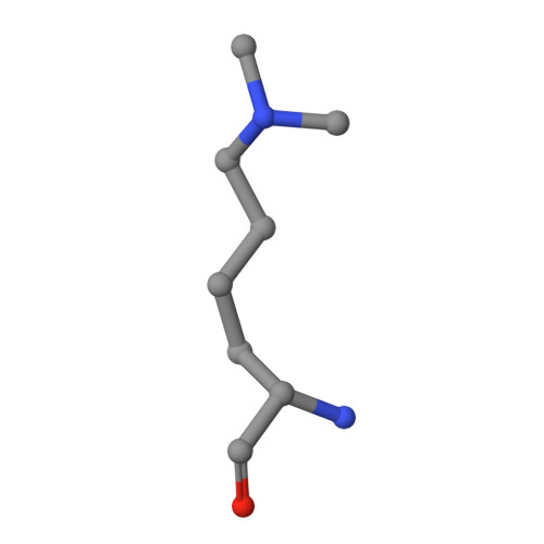
    The tumor suppressor p53 and the DNA repair factor 53BP1 (p53 binding protein 1) regulate gene transcription and responses to genotoxic stresses. Upon DNA damage, p53 undergoes dimethylation at Lys382 (p53K382me2), and this posttranslational modification is recognized by 53BP1. The molecular mechanism of nonhistone methyl-lysine mark recognition remains unknown. Here we report a 1. 6-A-resolution crystal structure of the tandem Tudor domain of human 53BP1 bound to a p53K382me2 peptide. In the complex, dimethylated Lys382 is restrained by a set of hydrophobic and cation-pi interactions in a cage formed by four aromatic residues and an aspartate of 53BP1. The signature HKKme2 motif of p53, which defines specificity, is identified through a combination of NMR resonance perturbations, mutagenesis, measurements of binding affinities and docking simulations, and analysis of the crystal structures of 53BP1 bound to p53 peptides containing other dimethyl-lysine marks, p53K370me2 (p53 dimethylated at Lys370) and p53K372me2 (p53 dimethylated at Lys372). Binding of the 53BP1 Tudor domain to p53K382me2 may facilitate p53 accumulation at DNA damage sites and promote DNA repair as suggested by chromatin immunoprecipitation and DNA repair assays. Together, our data detail the molecular mechanism of p53-53BP1 association and provide the basis for deciphering the role of this interaction in the regulation of p53 and 53BP1 functions.


  • Organizational Affiliation
    • Department of Pharmacology, University of Colorado Denver School of Medicine, 12801 East 17th Avenue, Aurora, CO 80045-0511, USA.

Macromolecule Content 

  • Total Structure Weight: 15.56 kDa 
  • Atom Count: 1,181 
  • Modeled Residue Count: 120 
  • Deposited Residue Count: 135 
  • Unique protein chains: 2

Macromolecules

Find similar proteins by:|  3D Structure
Entity ID: 1
MoleculeChains  Sequence LengthOrganismDetailsImage
Tumor suppressor p53-binding protein 1125Homo sapiensMutation(s): 0 
Gene Names: TP53BP1
UniProt & NIH Common Fund Data Resources
Find proteins for Q12888 (Homo sapiens)
Explore Q12888 
Go to UniProtKB:  Q12888
PHAROS:  Q12888
GTEx:  ENSG00000067369 
Entity Groups
Sequence Clusters30% Identity50% Identity70% Identity90% Identity95% Identity100% Identity
UniProt GroupQ12888
Sequence Annotations
Expand
Reference Sequence
Find similar proteins by:  Sequence   |   3D Structure  
Entity ID: 2
MoleculeChains  Sequence LengthOrganismDetailsImage
DIMETHYLATED p53 Lysine 370 PEPTIDE10N/AMutation(s): 0 

Experimental Data & Validation

Experimental Data

  • Method: X-RAY DIFFRACTION
  • Resolution: 1.50 Å
  • R-Value Free:  0.232 (Depositor), 0.230 (DCC) 
  • R-Value Work:  0.217 (Depositor), 0.212 (DCC) 
  • R-Value Observed: 0.217 (Depositor) 
Space Group: C 1 2 1
Unit Cell:
Length ( Å )Angle ( ˚ )
a = 56.44α = 90
b = 78.1β = 121.92
c = 36.31γ = 90
Software Package:
Software NamePurpose
CrystalCleardata collection
PHASERphasing
PHENIXrefinement
d*TREKdata reduction
d*TREKdata scaling

Structure Validation

View Full Validation Report


Entry History 

Deposition Data

Revision History  (Full details and data files)

  • Version 1.0: 2010-03-23
    Type: Initial release
  • Version 1.1: 2011-07-13
    Changes: Version format compliance
  • Version 1.2: 2023-09-06
    Changes: Data collection, Database references, Derived calculations, Refinement description