


Mechanism of Action (and Inhibition) of Head-to-Head Terpene Synthases: A Structural Investigation
Lin, F.-Y., Liu, C.-I., Liu, Y.-L., Zhang, Y., Wang, K., Cao, R., Wang, A.H.J., Oldfield, E.To be published.
Experimental Data Snapshot
Macromolecule Content
Entity ID: 1 | |||||
|---|---|---|---|---|---|
| Molecule | Chains | Sequence Length | Organism | Details | Image |
| Squalene synthetase | 340 | Homo sapiens | Mutation(s): 0 Gene Names: FDFT1 EC: 2.5.1.21 | 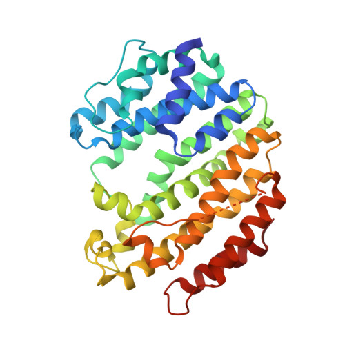 | |
UniProt & NIH Common Fund Data Resources | |||||
PHAROS: P37268 GTEx: ENSG00000079459 | |||||
Entity Groups | |||||
| Sequence Clusters | 30% Identity50% Identity70% Identity90% Identity95% Identity100% Identity | ||||
| UniProt Group | P37268 | ||||
Sequence AnnotationsExpand | |||||
Reference Sequence | |||||
| Ligands 2 Unique | |||||
|---|---|---|---|---|---|
| ID | Chains | Name / Formula / InChI Key | 2D Diagram | 3D Interactions | |
| B65 Download:Ideal Coordinates CCD File | G [auth A] I [auth B] K [auth C] M [auth D] O [auth E] | (1R)-4-(3-phenoxyphenyl)-1-phosphonobutane-1-sulfonic acid C16 H19 O7 P S RCGCZPXSRLLKCK-MRXNPFEDSA-N |  | ||
| MG Download:Ideal Coordinates CCD File | H [auth A] J [auth B] L [auth C] N [auth D] P [auth E] | MAGNESIUM ION Mg JLVVSXFLKOJNIY-UHFFFAOYSA-N |  | ||
| Length ( Å ) | Angle ( ˚ ) |
|---|---|
| a = 85.662 | α = 90 |
| b = 153.422 | β = 90.5 |
| c = 92.904 | γ = 90 |
| Software Name | Purpose |
|---|---|
| HKL-2000 | data collection |
| PHASES | phasing |
| REFMAC | refinement |
| HKL-2000 | data reduction |
| HKL-2000 | data scaling |


