3LE4 | pdb_00003le4

Crystal structure of the DGCR8 dimerization domain


Experimental Data Snapshot

  • Method: X-RAY DIFFRACTION
  • Resolution: 1.70 Å
  • R-Value Free: 
    0.213 (Depositor), 0.211 (DCC) 
  • R-Value Work: 
    0.181 (Depositor), 0.179 (DCC) 
  • R-Value Observed: 
    0.182 (Depositor) 

wwPDB Validation 3D Report Full Report

Validation slider image for 3LE4

This is version 1.2 of the entry. See complete history

Literature

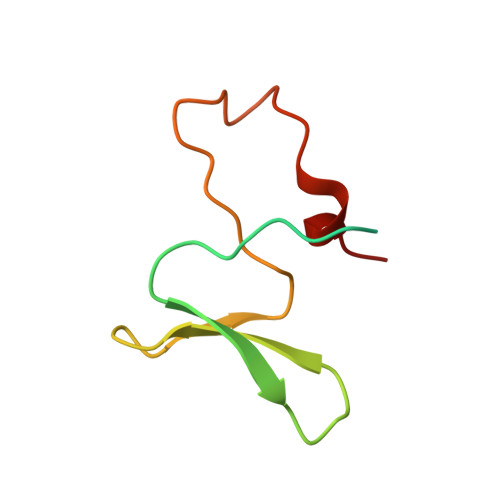
Structure of the dimerization domain of DiGeorge Critical Region 8

Senturia, R.Faller, M.Yin, S.Loo, J.A.Cascio, D.Sawaya, M.R.Hwang, D.Clubb, R.T.Guo, F.

(2010) Protein Sci 19: 1354-1365

  • DOI: https://doi.org/10.1002/pro.414
  • Primary Citation Related Structures: 
    3LE4

  • PubMed Abstract: 

    Maturation of microRNAs (miRNAs, approximately 22nt) from long primary transcripts [primary miRNAs (pri-miRNAs)] is regulated during development and is altered in diseases such as cancer. The first processing step is a cleavage mediated by the Microprocessor complex containing the Drosha nuclease and the RNA-binding protein DiGeorge critical region 8 (DGCR8). We previously reported that dimeric DGCR8 binds heme and that the heme-bound DGCR8 is more active than the heme-free form. Here, we identified a conserved dimerization domain in DGCR8. Our crystal structure of this domain (residues 298-352) at 1.7 A resolution demonstrates a previously unknown use of a WW motif as a platform for extensive dimerization interactions. The dimerization domain of DGCR8 is embedded in an independently folded heme-binding domain and directly contributes to association with heme. Heme-binding-deficient DGCR8 mutants have reduced pri-miRNA processing activity in vitro. Our study provides structural and biochemical bases for understanding how dimerization and heme binding of DGCR8 may contribute to regulation of miRNA biogenesis.


  • Organizational Affiliation
    • Department of Biological Chemistry in David Geffen School of Medicine, University of California, Los Angeles, California 90095, USA.

Macromolecule Content 

  • Total Structure Weight: 8.89 kDa 
  • Atom Count: 476 
  • Modeled Residue Count: 55 
  • Deposited Residue Count: 79 
  • Unique protein chains: 1

Macromolecules

Find similar proteins by:|  3D Structure
Entity ID: 1
MoleculeChains  Sequence LengthOrganismDetailsImage
Microprocessor complex subunit DGCR879Homo sapiensMutation(s): 0 
Gene Names: DGCR8C22orf12DGCRK6LP4941
UniProt & NIH Common Fund Data Resources
Find proteins for Q8WYQ5 (Homo sapiens)
Explore Q8WYQ5 
Go to UniProtKB:  Q8WYQ5
PHAROS:  Q8WYQ5
GTEx:  ENSG00000128191 
Entity Groups
Sequence Clusters30% Identity50% Identity70% Identity90% Identity95% Identity100% Identity
UniProt GroupQ8WYQ5
Sequence Annotations
Expand
Reference Sequence

Experimental Data & Validation

Experimental Data

  • Method: X-RAY DIFFRACTION
  • Resolution: 1.70 Å
  • R-Value Free:  0.213 (Depositor), 0.211 (DCC) 
  • R-Value Work:  0.181 (Depositor), 0.179 (DCC) 
  • R-Value Observed: 0.182 (Depositor) 
Space Group: P 43 21 2
Unit Cell:
Length ( Å )Angle ( ˚ )
a = 39.952α = 90
b = 39.952β = 90
c = 80.626γ = 90
Software Package:
Software NamePurpose
ADSCdata collection
SHELXSphasing
PHENIXrefinement
HKL-2000data reduction
HKL-2000data scaling

Structure Validation

View Full Validation Report


Entry History 

Deposition Data

Revision History  (Full details and data files)

  • Version 1.0: 2010-07-07
    Type: Initial release
  • Version 1.1: 2011-07-13
    Changes: Version format compliance
  • Version 1.2: 2024-02-21
    Changes: Data collection, Database references