3LDY

An extraordinary mechanism of DNA perturbation exhibited by the rare-cutting HNH restriction endonuclease PacI


Experimental Data Snapshot

  • Method: X-RAY DIFFRACTION
  • Resolution: 1.97 Å
  • R-Value Free: 0.217 
  • R-Value Work: 0.184 
  • R-Value Observed: 0.186 

wwPDB Validation   3D Report Full Report


This is version 1.4 of the entry. See complete history


Literature

Unusual target site disruption by the rare-cutting HNH restriction endonuclease PacI.

Shen, B.W.Heiter, D.F.Chan, S.H.Wang, H.Xu, S.Y.Morgan, R.D.Wilson, G.G.Stoddard, B.L.

(2010) Structure 18: 734-743

  • DOI: https://doi.org/10.1016/j.str.2010.03.009
  • Primary Citation of Related Structures:  
    3LDY, 3M7K

  • PubMed Abstract: 

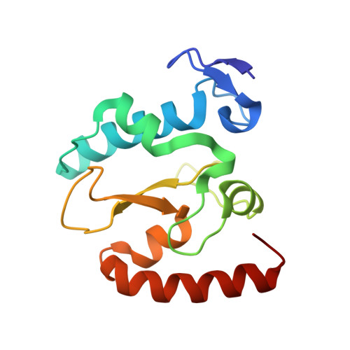
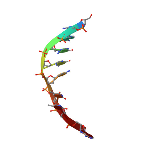
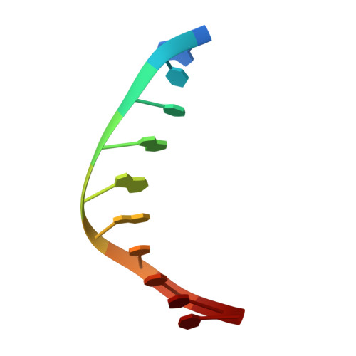
    The crystal structure of the rare-cutting HNH restriction endonuclease PacI in complex with its eight-base-pair target recognition sequence 5'-TTAATTAA-3' has been determined to 1.9 A resolution. The enzyme forms an extended homodimer, with each subunit containing two zinc-bound motifs surrounding a betabetaalpha-metal catalytic site. The latter is unusual in that a tyrosine residue likely initiates strand cleavage. PacI dramatically distorts its target sequence from Watson-Crick duplex DNA base pairing, with every base separated from its original partner. Two bases on each strand are unpaired, four are engaged in noncanonical A:A and T:T base pairs, and the remaining two bases are matched with new Watson-Crick partners. This represents a highly unusual DNA binding mechanism for a restriction endonuclease, and implies that initial recognition of the target site might involve significantly different contacts from those visualized in the DNA-bound cocrystal structures.


  • Organizational Affiliation

    Division of Basic Sciences, Fred Hutchinson Cancer Research Center, 1100 Fairview Avenue N. A3-025, Seattle, WA 98109, USA.


Macromolecules

Find similar proteins by:  (by identity cutoff)  |  3D Structure
Entity ID: 1
MoleculeChains Sequence LengthOrganismDetailsImage
restriction endonuclease PacI142Pseudomonas alcaligenesMutation(s): 0 
Gene Names: pacIR
UniProt
Find proteins for D5MNX7 (Pseudomonas alcaligenes)
Explore D5MNX7 
Go to UniProtKB:  D5MNX7
Entity Groups  
Sequence Clusters30% Identity50% Identity70% Identity90% Identity95% Identity100% Identity
UniProt GroupD5MNX7
Sequence Annotations
Expand
  • Reference Sequence

Find similar nucleic acids by:  Sequence   |   3D Structure  

Entity ID: 2
MoleculeChains LengthOrganismImage
DNA (5'-D(*GP*AP*GP*GP*CP*TP*TP*A)-3')8N/A
Sequence Annotations
Expand
  • Reference Sequence

Find similar nucleic acids by:  Sequence   |   3D Structure  

Entity ID: 3
MoleculeChains LengthOrganismImage
DNA (5'-D(P*AP*TP*TP*AP*AP*GP*CP*CP*TP*C)-3')10N/A
Sequence Annotations
Expand
  • Reference Sequence
Experimental Data & Validation

Experimental Data

  • Method: X-RAY DIFFRACTION
  • Resolution: 1.97 Å
  • R-Value Free: 0.217 
  • R-Value Work: 0.184 
  • R-Value Observed: 0.186 
  • Space Group: C 2 2 21
Unit Cell:
Length ( Å )Angle ( ˚ )
a = 36.908α = 90
b = 115.788β = 90
c = 114.689γ = 90
Software Package:
Software NamePurpose
ADSCdata collection
SOLVEphasing
REFMACrefinement
HKL-2000data reduction
SCALEPACKdata scaling

Structure Validation

View Full Validation Report


Entry History 

Deposition Data

Revision History  (Full details and data files)

  • Version 1.0: 2010-04-21
    Type: Initial release
  • Version 1.1: 2011-07-13
    Changes: Version format compliance
  • Version 1.2: 2011-07-20
    Changes: Database references
  • Version 1.3: 2013-07-17
    Changes: Refinement description
  • Version 1.4: 2024-02-21
    Changes: Data collection, Database references, Derived calculations