


The Crystal Structure of smu.1229 from Streptococcus mutans UA159 bound to hypoxanthine
Su, X.-D., Hou, Q.M., Wang, H.F., Liu, X.To be published.
Experimental Data Snapshot
wwPDB Validation 3D Report Full Report

Entity ID: 1 | |||||
|---|---|---|---|---|---|
| Molecule | Chains | Sequence Length | Organism | Details | Image |
| Putative purine nucleoside phosphorylase | 303 | Streptococcus mutans UA159 | Mutation(s): 0 Gene Names: smu.1229 EC: 2.4.2.1 | 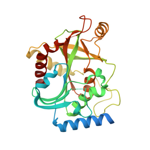 | |
UniProt | |||||
Entity Groups | |||||
| Sequence Clusters | 30% Identity50% Identity70% Identity90% Identity95% Identity100% Identity | ||||
| UniProt Group | Q8DTU4 | ||||
Sequence AnnotationsExpand | |||||
Reference Sequence | |||||
| Ligands 2 Unique | |||||
|---|---|---|---|---|---|
| ID | Chains | Name / Formula / InChI Key | 2D Diagram | 3D Interactions | |
| HPA Download:Ideal Coordinates CCD File | B [auth A] | HYPOXANTHINE C5 H4 N4 O FDGQSTZJBFJUBT-UHFFFAOYSA-N |  | ||
| SO4 Download:Ideal Coordinates CCD File | C [auth A] | SULFATE ION O4 S QAOWNCQODCNURD-UHFFFAOYSA-L |  | ||
| Length ( Å ) | Angle ( ˚ ) |
|---|---|
| a = 83.5 | α = 90 |
| b = 83.5 | β = 90 |
| c = 127.76 | γ = 120 |
| Software Name | Purpose |
|---|---|
| ADSC | data collection |
| MOLREP | phasing |
| CNS | refinement |
| MOSFLM | data reduction |
| SCALA | data scaling |
| REFMAC | refinement |


