3L7V | pdb_00003l7v

Crystal structure of a hypothetical protein smu.1377c from Streptococcus mutans UA159


Experimental Data Snapshot

  • Method: X-RAY DIFFRACTION
  • Resolution: 2.26 Å
  • R-Value Free: 
    0.259 (Depositor), 0.183 (DCC) 
  • R-Value Work: 
    0.196 (Depositor), 0.196 (DCC) 
  • R-Value Observed: 
    0.199 (Depositor) 

wwPDB Validation 3D Report Full Report

Validation slider image for 3L7V

This is version 1.3 of the entry. See complete history

Literature

The structure of the hypothetical protein smu.1377c from Streptococcus mutans suggests a role in tRNA modification

Fu, T.-M.Liu, X.Li, L.Su, X.-D.

(2010) Acta Crystallogr Sect F Struct Biol Cryst Commun 66: 771-775

  • DOI: https://doi.org/10.1107/S1744309110018944
  • Primary Citation Related Structures: 
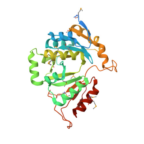
    3L7V

  • PubMed Abstract: 

    Members of the Sua5_YciO_YrdC protein family are found in both eukaryotes and prokaryotes and possess a conserved alpha/beta twisted open-sheet fold. The Escherichia coli protein YrdC has been shown to be involved in modification of tRNA. The crystal structure of smu.1377c, a hypothetical protein from Streptococcus mutans, has been determined to 2.25 A resolution. From structure analysis and comparison, it is shown that smu.1377c is a member of the Sua5_YciO_YrdC family and that it may play the same role as E. coli YrdC.


  • Organizational Affiliation
    • The National Laboratory of Protein Engineering and Plant Genetic Engineering, School of Life Sciences, Peking University, Beijing 100871, People's Republic of China.

Macromolecule Content 

  • Total Structure Weight: 34.41 kDa 
  • Atom Count: 2,127 
  • Modeled Residue Count: 256 
  • Deposited Residue Count: 295 
  • Unique protein chains: 1

Macromolecules

Find similar proteins by:|  3D Structure
Entity ID: 1
MoleculeChains  Sequence LengthOrganismDetailsImage
Putative uncharacterized protein smu.1377c295Streptococcus mutans UA159Mutation(s): 0 
Gene Names: smu.1377c
UniProt
Find proteins for Q8DTG7 (Streptococcus mutans serotype c (strain ATCC 700610 / UA159))
Explore Q8DTG7 
Go to UniProtKB:  Q8DTG7
Entity Groups
Sequence Clusters30% Identity50% Identity70% Identity90% Identity95% Identity100% Identity
UniProt GroupQ8DTG7
Sequence Annotations
Expand
Reference Sequence

Experimental Data & Validation

Experimental Data

  • Method: X-RAY DIFFRACTION
  • Resolution: 2.26 Å
  • R-Value Free:  0.259 (Depositor), 0.183 (DCC) 
  • R-Value Work:  0.196 (Depositor), 0.196 (DCC) 
  • R-Value Observed: 0.199 (Depositor) 
Space Group: P 21 21 2
Unit Cell:
Length ( Å )Angle ( ˚ )
a = 81.9α = 90
b = 92.6β = 90
c = 40.6γ = 90
Software Package:
Software NamePurpose
MAR345dtbdata collection
SHELXSphasing
PHENIXrefinement
XDSdata reduction
XSCALEdata scaling

Structure Validation

View Full Validation Report


Entry History 

Deposition Data

Revision History  (Full details and data files)

  • Version 1.0: 2010-07-21
    Type: Initial release
  • Version 1.1: 2011-07-13
    Changes: Version format compliance
  • Version 1.2: 2018-05-30
    Changes: Data collection
  • Version 1.3: 2024-10-30
    Changes: Data collection, Database references, Derived calculations, Structure summary