Cysteine as a modulator residue in the active site of xenobiotic reductase A: a structural, thermodynamic and kinetic study
Spiegelhauer, O., Mende, S., Dickert, F., Knauer, S.H., Ullmann, G.M., Dobbek, H.(2010) J Mol Biol 398: 66-82
- PubMed: 20206186 Search on PubMed
- DOI: https://doi.org/10.1016/j.jmb.2010.02.044
- Primary Citation Related Structures:
3L5L, 3L5M, 3L65, 3L66, 3L67, 3L68 - PubMed Abstract:
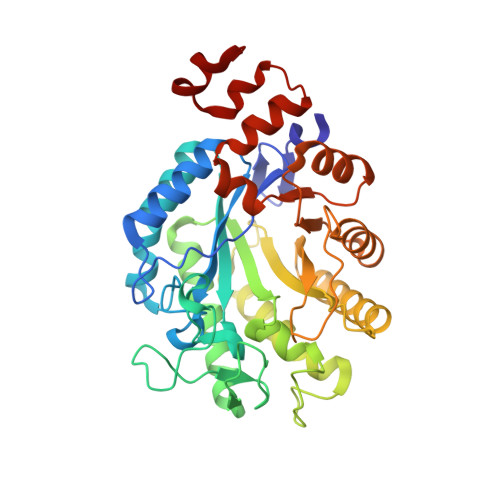
Xenobiotic reductase A (XenA) from Pseudomonas putida 86 catalyzes the NADH/NADPH-dependent reduction of various substrates, including 2-cyclohexenone and 8-hydroxycoumarin. XenA is a member of the old yellow enzyme (OYE) family of flavoproteins and is structurally and functionally similar to other bacterial members of this enzyme class. A characteristic feature of XenA is the presence of a cysteine residue (Cys25) in the active site, where in most members of the OYE family a threonine residue is found that modulates the reduction potential of the FMN/FMNH(-) couple. We investigated the role of Cys25 by studying two variants in which the residue has been exchanged for a serine and an alanine residue. While the exchange against alanine has a remarkably small effect on the reduction potential, the reactivity and the structure of XenA, the exchange against serine increases the reduction potential by +82 mV, increases the rate constant of the reductive half-reaction and decreases the rate constant in the oxidative half-reaction. We determined six crystal structures at high to true atomic resolution (d(min) 1.03-1.80 A) of the three XenA variants with and without the substrate coumarin bound in the active site. The atomic resolution structure of XenA in complex with coumarin reveals a compressed active site geometry in which the isoalloxazine ring is sandwiched between coumarin and the protein backbone. The structures further reveal that the conformation of the active site and substrate interactions are preserved in the two variants, indicating that the observed changes are due to local effects only. We propose that Cys25 and the residues in its place determine which of the two half-reactions is rate limiting, depending on the substrate couple. This might help to explain why the genome of Pseudomonas putida encodes multiple xenobiotic reductases containing either cysteine, threonine or alanine in the active site.
- Bioinorganic Chemistry, Universität Bayreuth, Universitätsstrasse 30, 95447 Bayreuth, Germany.
Organizational Affiliation: