3L51

Crystal Structure of the Mouse Condensin Hinge Domain


Experimental Data Snapshot

  • Method: X-RAY DIFFRACTION
  • Resolution: 1.51 Å
  • R-Value Free: 0.173 
  • R-Value Work: 0.143 
  • R-Value Observed: 0.146 

wwPDB Validation   3D Report Full Report


This is version 1.1 of the entry. See complete history


Literature

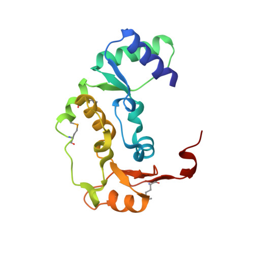
Structure and DNA binding activity of the mouse condensin hinge domain highlight common and diverse features of SMC proteins

Griese, J.J.Witte, G.Hopfner, K.-P.

(2010) Nucleic Acids Res 38: 3454-3465

  • DOI: https://doi.org/10.1093/nar/gkq038
  • Primary Citation of Related Structures:  
    3L51

  • PubMed Abstract: 

    Structural Maintenance of Chromosomes (SMC) proteins are vital for a wide range of processes including chromosome structure and dynamics, gene regulation and DNA repair. Eukaryotes have three SMC complexes, consisting of heterodimeric pairs of six different SMC proteins along with several specific regulatory subunits. In addition to their other functions, all three SMC complexes play distinct roles in DNA repair. Cohesin (SMC1-SMC3) is involved in DNA double-strand break repair, condensin (SMC2-SMC4) participates in single-strand break (SSB) repair, and the SMC5-SMC6 complex functions in various DNA repair pathways. SMC proteins consist of N- and C-terminal domains that fold back onto each other to create an ATPase 'head' domain, connected to a central 'hinge' domain via long coiled-coils. The hinge domain mediates dimerization of SMC proteins and binds DNA, but it is not clear to what purpose this activity serves. We studied the structure and function of the condensin hinge domain from mouse. While the SMC hinge domain structure is largely conserved from prokaryotes to eukaryotes, its function seems to have diversified throughout the course of evolution. The condensin hinge domain preferentially binds single-stranded DNA. We propose that this activity plays a role in the SSB repair function of the condensin complex.


  • Organizational Affiliation

    Department of Biochemistry, Gene Center, Center for Integrated Protein Sciences and Munich Center for Advanced Photonics, Ludwig-Maximilians University Munich, Feodor-Lynen-Str 25, D-81377 Munich, Germany.


Macromolecules
Find similar proteins by:  (by identity cutoff)  |  3D Structure
Entity ID: 1
MoleculeChains Sequence LengthOrganismDetailsImage
Structural maintenance of chromosomes protein 2161Mus musculusMutation(s): 0 
Gene Names: SMC2 (amino acids 506-666)
UniProt
Find proteins for Q8CG48 (Mus musculus)
Explore Q8CG48 
Go to UniProtKB:  Q8CG48
Entity Groups  
Sequence Clusters30% Identity50% Identity70% Identity90% Identity95% Identity100% Identity
UniProt GroupQ8CG48
Sequence Annotations
Expand
  • Reference Sequence
Find similar proteins by:  (by identity cutoff)  |  3D Structure
Entity ID: 2
MoleculeChains Sequence LengthOrganismDetailsImage
Structural maintenance of chromosomes protein 4166Mus musculusMutation(s): 0 
Gene Names: SMC4 (amino acids 595-752)
UniProt
Find proteins for Q8CG47 (Mus musculus)
Explore Q8CG47 
Go to UniProtKB:  Q8CG47
Entity Groups  
Sequence Clusters30% Identity50% Identity70% Identity90% Identity95% Identity100% Identity
UniProt GroupQ8CG47
Sequence Annotations
Expand
  • Reference Sequence
Experimental Data & Validation

Experimental Data

  • Method: X-RAY DIFFRACTION
  • Resolution: 1.51 Å
  • R-Value Free: 0.173 
  • R-Value Work: 0.143 
  • R-Value Observed: 0.146 
  • Space Group: P 1 21 1
Unit Cell:
Length ( Å )Angle ( ˚ )
a = 33.455α = 90
b = 96.741β = 92.84
c = 54.537γ = 90
Software Package:
Software NamePurpose
XSCALEdata scaling
SHARPphasing
DMphasing
PHENIXrefinement
PDB_EXTRACTdata extraction
XDSdata reduction
XDSdata scaling

Structure Validation

View Full Validation Report


Entry History 

Deposition Data

Revision History  (Full details and data files)

  • Version 1.0: 2010-02-16
    Type: Initial release
  • Version 1.1: 2011-07-13
    Changes: Version format compliance