3L4M

Crystal Structure of the MauG/pre-Methylamine Dehydrogenase Complex.


Experimental Data Snapshot

  • Method: X-RAY DIFFRACTION
  • Resolution: 2.02 Å
  • R-Value Free: 0.189 
  • R-Value Work: 0.135 
  • R-Value Observed: 0.138 

wwPDB Validation   3D Report Full Report


Ligand Structure Quality Assessment 


This is version 1.4 of the entry. See complete history


Literature

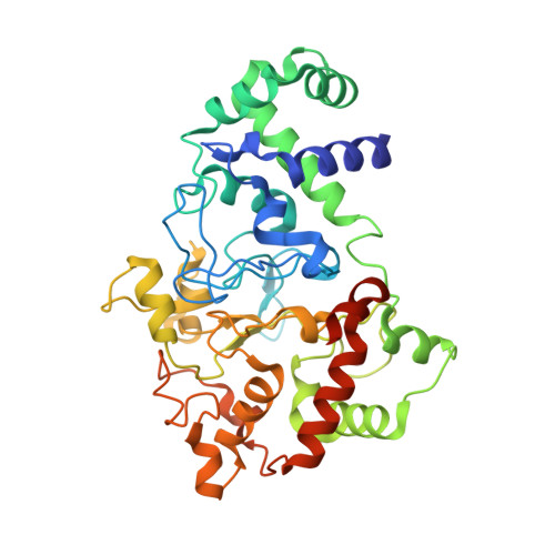
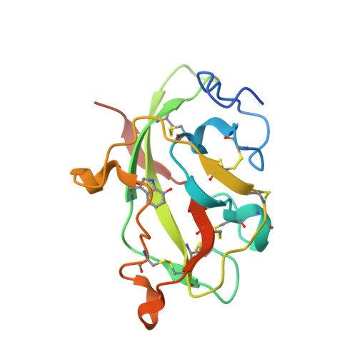
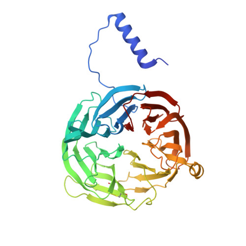
In crystallo posttranslational modification within a MauG/pre-methylamine dehydrogenase complex.

Jensen, L.M.Sanishvili, R.Davidson, V.L.Wilmot, C.M.

(2010) Science 327: 1392-1394

  • DOI: https://doi.org/10.1126/science.1182492
  • Primary Citation of Related Structures:  
    3L4M, 3L4O

  • PubMed Abstract: 

    MauG is a diheme enzyme responsible for the posttranslational modification of two tryptophan residues to form the tryptophan tryptophylquinone (TTQ) cofactor of methylamine dehydrogenase (MADH). MauG converts preMADH, containing monohydroxylated betaTrp57, to fully functional MADH by catalyzing the insertion of a second oxygen atom into the indole ring and covalently linking betaTrp57 to betaTrp108. We have solved the x-ray crystal structure of MauG complexed with preMADH to 2.1 angstroms. The c-type heme irons and the nascent TTQ site are separated by long distances over which electron transfer must occur to achieve catalysis. In addition, one of the hemes has an atypical His-Tyr axial ligation. The crystalline protein complex is catalytically competent; upon addition of hydrogen peroxide, MauG-dependent TTQ synthesis occurs.


  • Organizational Affiliation

    Department of Biochemistry, Molecular Biology and Biophysics, University of Minnesota, Minneapolis, MN 55455, USA.


Macromolecules
Find similar proteins by:  (by identity cutoff)  |  3D Structure
Entity ID: 1
MoleculeChains Sequence LengthOrganismDetailsImage
Methylamine utilization protein mauG
A, B
373Paracoccus denitrificans PD1222Mutation(s): 0 
Gene Names: mauG
EC: 1
UniProt
Find proteins for Q51658 (Paracoccus denitrificans (strain Pd 1222))
Explore Q51658 
Go to UniProtKB:  Q51658
Entity Groups  
Sequence Clusters30% Identity50% Identity70% Identity90% Identity95% Identity100% Identity
UniProt GroupQ51658
Sequence Annotations
Expand
  • Reference Sequence
Find similar proteins by:  (by identity cutoff)  |  3D Structure
Entity ID: 2
MoleculeChains Sequence LengthOrganismDetailsImage
Methylamine dehydrogenase light chain
C, E
137Paracoccus denitrificans PD1222Mutation(s): 0 
Gene Names: mauA
EC: 1.4.99.3
UniProt
Find proteins for A1BBA0 (Paracoccus denitrificans (strain Pd 1222))
Explore A1BBA0 
Go to UniProtKB:  A1BBA0
Entity Groups  
Sequence Clusters30% Identity50% Identity70% Identity90% Identity95% Identity100% Identity
UniProt GroupA1BBA0
Sequence Annotations
Expand
  • Reference Sequence
Find similar proteins by:  (by identity cutoff)  |  3D Structure
Entity ID: 3
MoleculeChains Sequence LengthOrganismDetailsImage
Methylamine dehydrogenase heavy chain
D, F
386Paracoccus denitrificans PD1222Mutation(s): 0 
Gene Names: Pden_4730
EC: 1.4.99.3
UniProt
Find proteins for A1BB97 (Paracoccus denitrificans (strain Pd 1222))
Explore A1BB97 
Go to UniProtKB:  A1BB97
Entity Groups  
Sequence Clusters30% Identity50% Identity70% Identity90% Identity95% Identity100% Identity
UniProt GroupA1BB97
Sequence Annotations
Expand
  • Reference Sequence
Small Molecules
Ligands 5 Unique
IDChains Name / Formula / InChI Key2D Diagram3D Interactions
HEC
Query on HEC

Download Ideal Coordinates CCD File 
H [auth A],
I [auth A],
K [auth B],
L [auth B]
HEME C
C34 H34 Fe N4 O4
HXQIYSLZKNYNMH-LJNAALQVSA-N
1PE
Query on 1PE

Download Ideal Coordinates CCD File 
M [auth F]PENTAETHYLENE GLYCOL
C10 H22 O6
JLFNLZLINWHATN-UHFFFAOYSA-N
PG4
Query on PG4

Download Ideal Coordinates CCD File 
N [auth F]TETRAETHYLENE GLYCOL
C8 H18 O5
UWHCKJMYHZGTIT-UHFFFAOYSA-N
ACT
Query on ACT

Download Ideal Coordinates CCD File 
O [auth F]ACETATE ION
C2 H3 O2
QTBSBXVTEAMEQO-UHFFFAOYSA-M
CA
Query on CA

Download Ideal Coordinates CCD File 
G [auth A],
J [auth B]
CALCIUM ION
Ca
BHPQYMZQTOCNFJ-UHFFFAOYSA-N
Modified Residues  1 Unique
IDChains TypeFormula2D DiagramParent
0AF
Query on 0AF
C, E
L-PEPTIDE LINKINGC11 H12 N2 O3TRP
Experimental Data & Validation

Experimental Data

  • Method: X-RAY DIFFRACTION
  • Resolution: 2.02 Å
  • R-Value Free: 0.189 
  • R-Value Work: 0.135 
  • R-Value Observed: 0.138 
  • Space Group: P 1
Unit Cell:
Length ( Å )Angle ( ˚ )
a = 55.527α = 109.94
b = 83.524β = 91.54
c = 107.782γ = 105.78
Software Package:
Software NamePurpose
DENZOdata reduction
SCALEPACKdata scaling
PHASERphasing
REFMACrefinement
PDB_EXTRACTdata extraction
Blu-Icedata collection

Structure Validation

View Full Validation Report


Ligand Structure Quality Assessment 


Entry History 

Deposition Data

Revision History  (Full details and data files)

  • Version 1.0: 2010-03-23
    Type: Initial release
  • Version 1.1: 2011-07-13
    Changes: Advisory, Refinement description, Version format compliance
  • Version 1.2: 2017-11-01
    Changes: Refinement description
  • Version 1.3: 2023-09-06
    Changes: Data collection, Database references, Derived calculations, Refinement description
  • Version 1.4: 2023-11-22
    Changes: Data collection